This Week in Dental Implants we are highlighting a guided bone regeneration case (GBR) case using DALI grafting materials. The patient presented with a poorly healed site with previously failed dental implants. An aggressive guided bone regeneration procedure was indicated to facilitate bone growth for future implant placement, including the use of DALI Amnion Chorion Membrane .
Case Presentation
Case Presented By: Dr. Daniel Gober, of Marine Park Perio
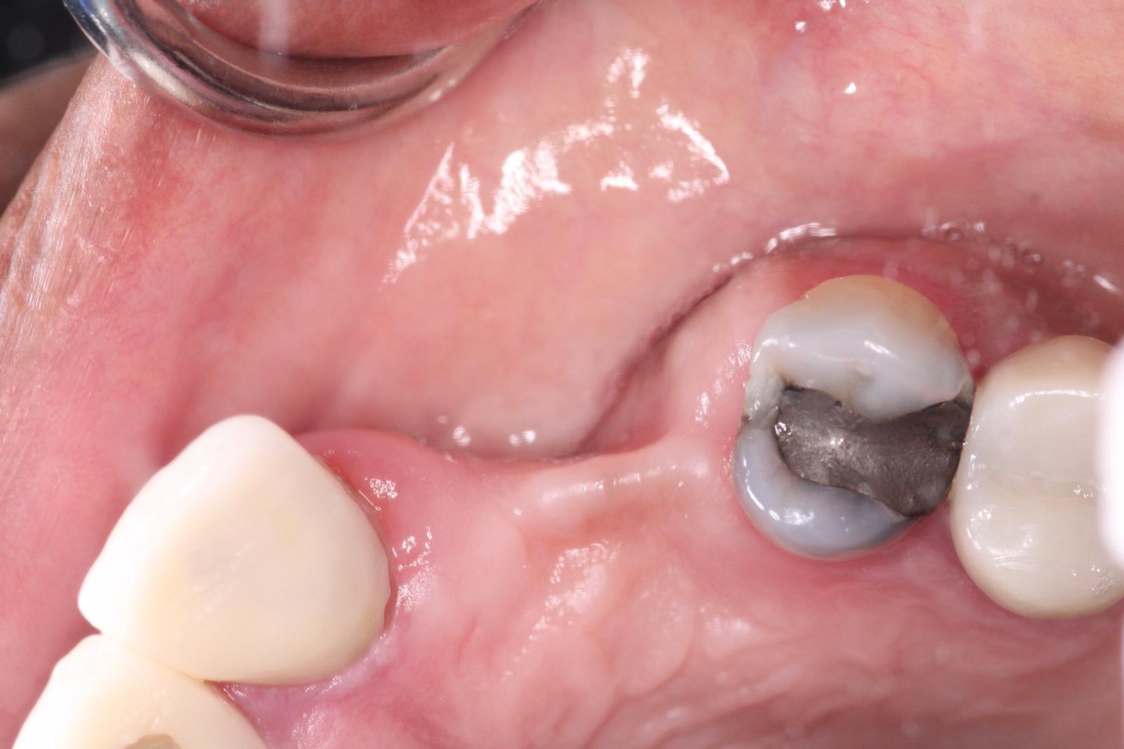
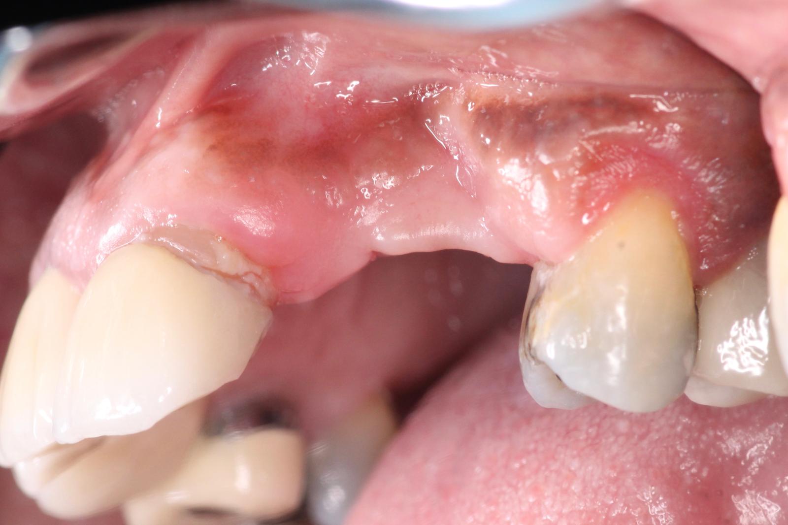
Missing 10 & 11 were extracted a year ago. Implants were placed, but healed terribly. The failed implants were removed, and now the site presents with significant bone loss, and severe resorption.
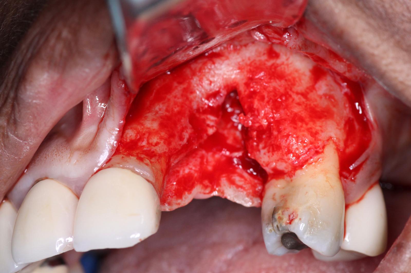
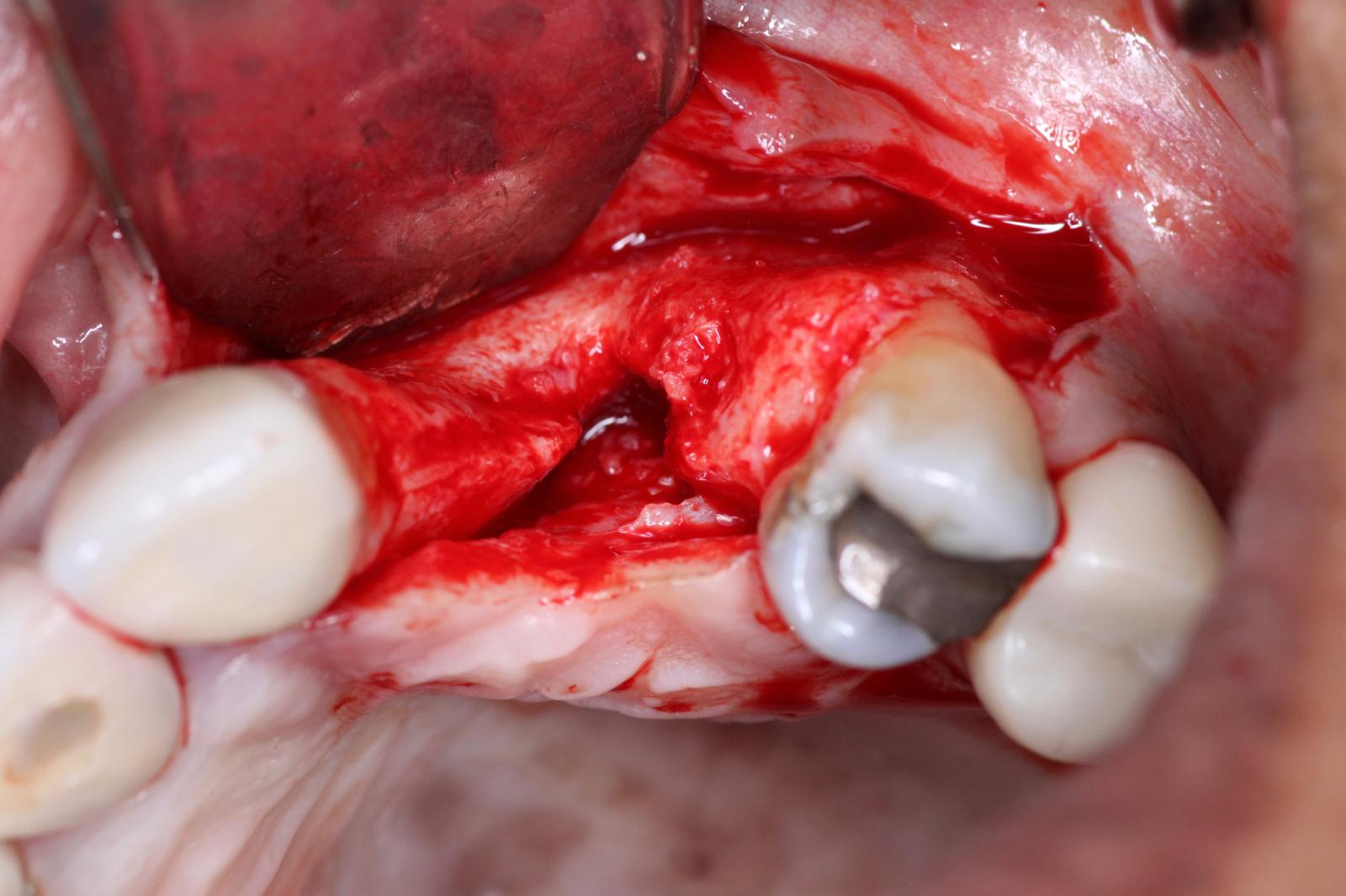
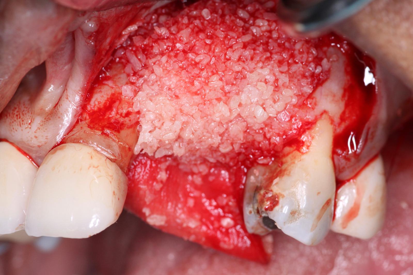
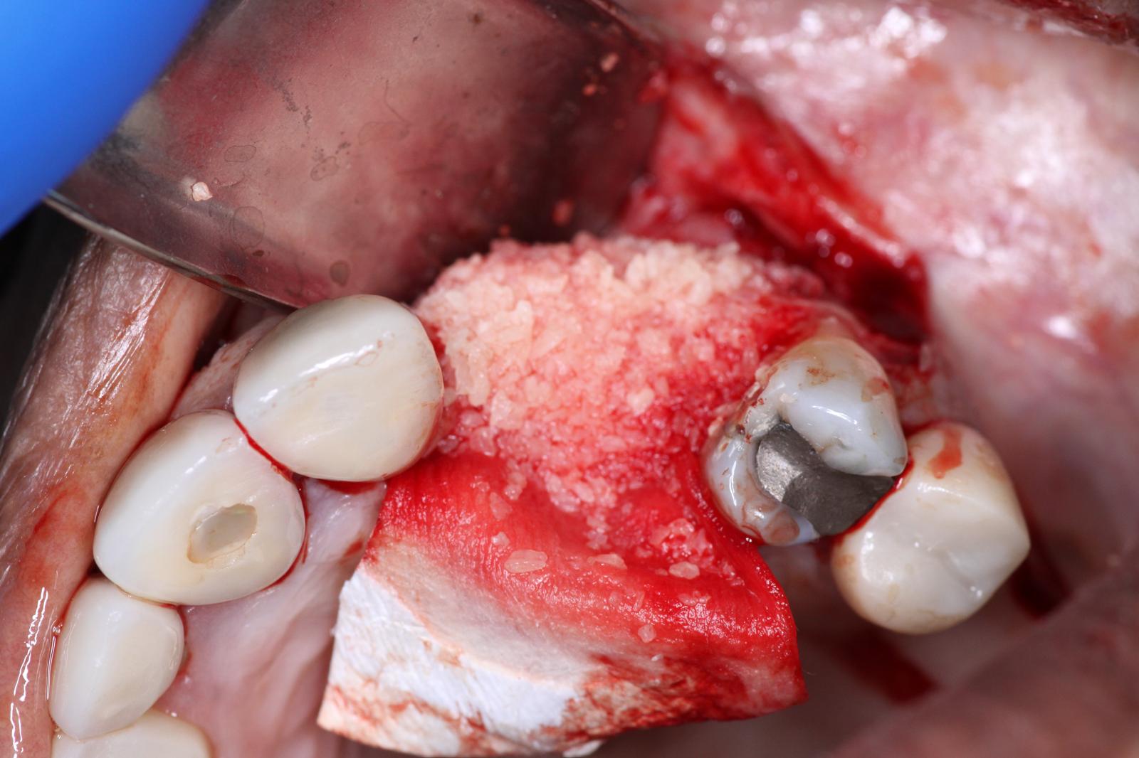
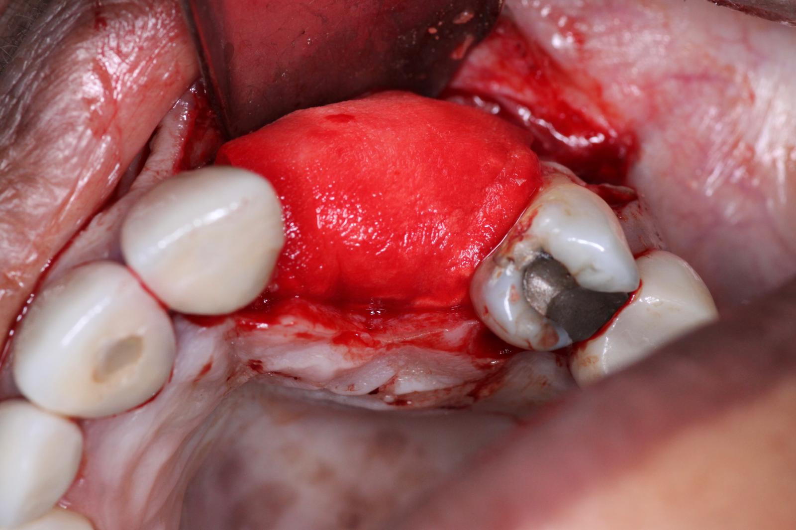
A Full thickness flap was reflected to expose the defect. You can see how severe the defect is, in both the vertical and horizontal dimensions. This is going to be a challenging GBR procedure, and we wanted to use the best materials at our disposal to encourage bone growth.
The first part of our grafting technique involved placement of DALI Cortical Cancellous Graft and initial placement of OsseoSeal Collagen Membrane. Notice how we position the membrane to tuck it under the palatal flap to help build up the bone graft. (for additional information on this technique see: Placement Techniques for OsseoSeal Porcine Collagen Membrane and Using OsseoSeal for Buccal Contour Augmentation )
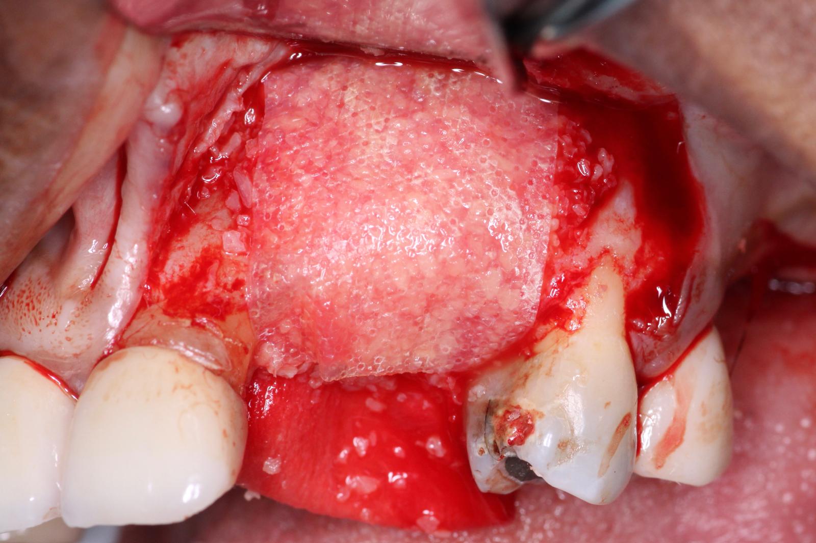
In the second part of our grafting technique, a DALI Amnion Chorion Membrane was layered over the bone graft, before we covered the entire site with a collagen membrane. The rationale for using an Amnion membrane here is that given the difficult nature of this GBR, we wanted to take advantage of the unique biological benefits of Amnion membranes, such as their inherent growth factors and anti-bacterial properties, to maximize the healing of the site and and enhance regeneration. For additional information on using Amnion membranes for this purpose, please see Enhance GBR and Improve Implant Surgical Outcomes with Amnion-Chorion Membranes
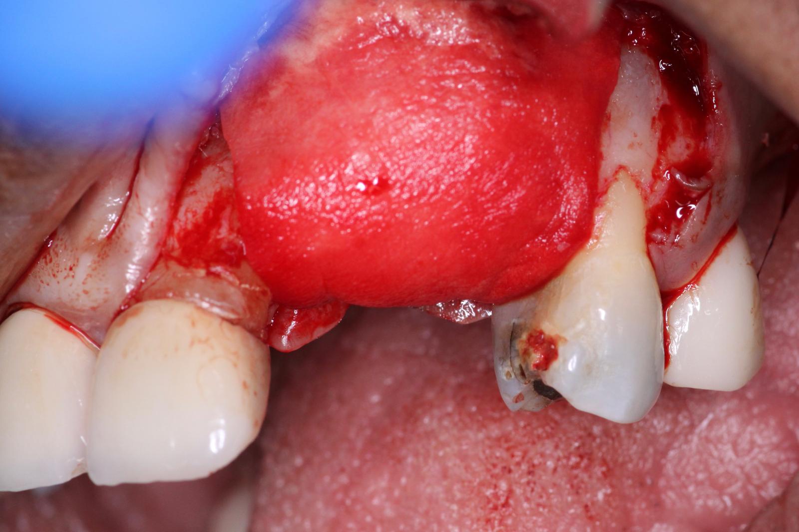
Next, we folded over the OsseoSeal collagen membrane to cover the bone and extend onto the native bone margins. It is crucial to try to cover everything and create a large enough space for the graft material to heal.
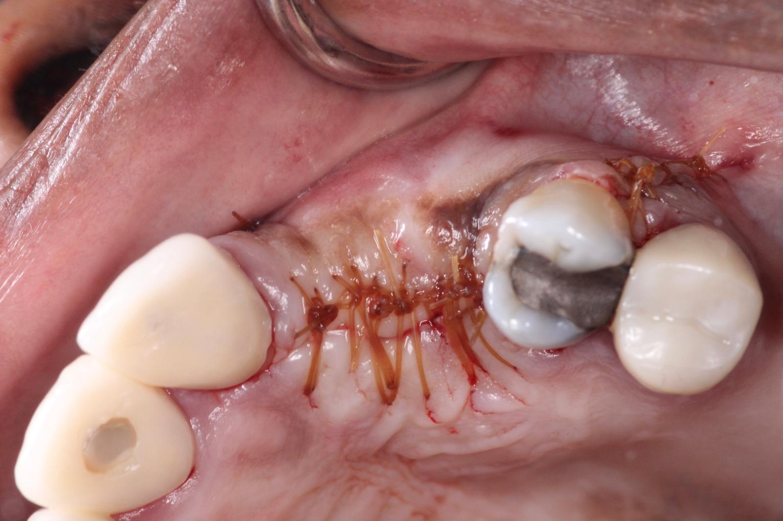
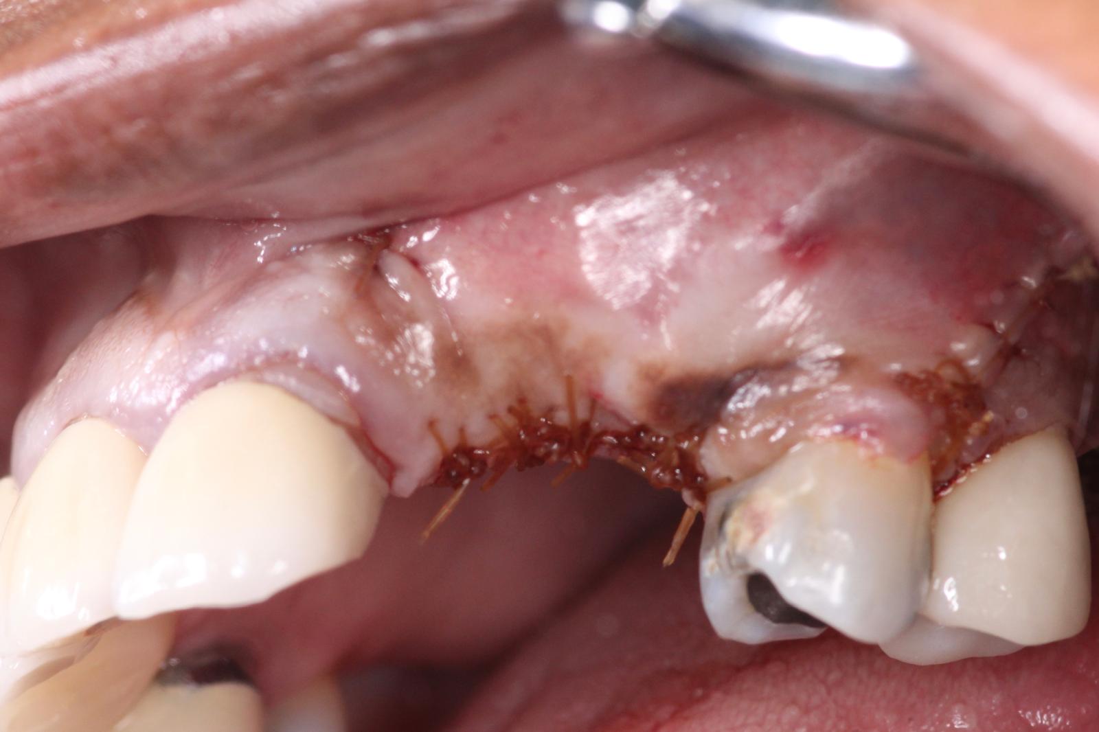
Finally, we sutured the site using Chromic Gut sutures. It is critical in GBR to get primary closure. In this particular case because of how much bone volume we are adding, a very deep periosteal release was done we we could pull over all the tissue from the buccal and suture the site as passively as possible. For more information this suturing technique, please see: Bone Regeneration of Extensive Socket Defects with Immediate Implant Placement
More to come…We will update this post in a few months with the post-operative results.