This Week in Dental Implants we wanted to share another case using a demineralized cortical allograft sheet in guided bone regeneration. Similar to our previous case, Grafting’s Best Kept Secret, the patient presented with a buccal ridge deficiency, which provided an excellent situation for consideration of a “flexible” deminearlized cortical sheet, like DALI Flex Graft. Because the demineralized cortical sheet is both rigid, and yet flexible, it allows clinicians to more easily create ideal ridge contours. Also, note the use of Cortical perforations in the case below. This topic is covered in a previous post, Cortical Bone Perforation in GBR: Is it necessary?
Case by: Vinay Bhide, DDS, MSc, FRCD of Aurora Periodontal Centre
In this case, Dr. Bhide shows how he used a demineralized cortical sheet, in tandem with GBR to restore a healthy 72-year-old patient’s missing tooth No. 9.
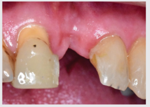
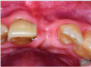
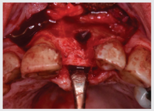
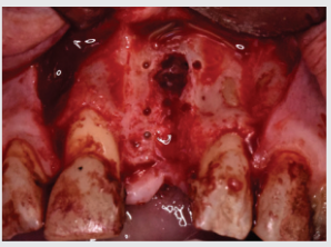
Figure 1: Patient presents with missing tooth No. 9 that was extracted 2 months earlier with uneventful healing.
Figure 2: Occlusal view showing obvious buccal alveolar ridge deficiency
Figure 3: Mucoperiosteal flap elevated to expose defect; note the bony dehiscence on the buccal aspect
Figure 4: Cortical perforations made in buccal bone
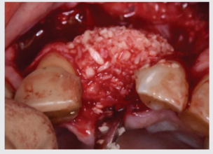
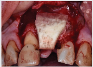
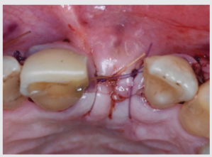
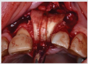
Figure 5: Deficient ridge grafted with mineralized cortico-cancellous particulate bone allograft.
Figure 6: Demineralized cortical sheet placed over particulate bone
Figure 7: Periosteal stabilizing suture placed to secure demineralized cortical sheet
Figure 8: Tension-free primary closure was achieved using resorbable monofilament sutures
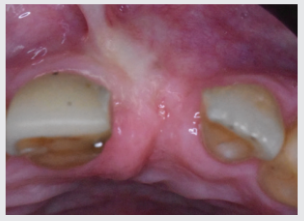
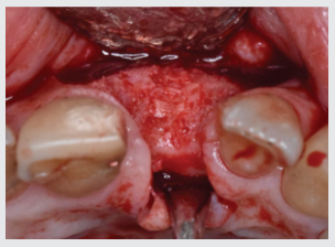
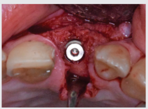
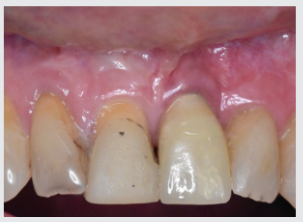
Figure 9: At 6 months, the grafted ridge has healed well and is now ready for implant placement surgery
Figure 10: Flap elevation reveals nicely reconstructed ridge with dimensions suitable for implant placement
Figure 11: Implant fixture of appropriate diameter and length was placed in grafted site with insertion torque valued of 35 Ncm; quality of grafted bone was D2.
Figure 12: The implant was restored with a screw-retained crown prosthesis 4 months after implant placement. This picture was taken 1 year following implant restoration.