Artificial intelligence (AI) is already revolutionizing many medical areas, but its application for implant dentistry is still not entirely clear. In this Week in Dental Implants, we start with a sample case demonstrating how AI is created and then tested for accuracy. Further down, we highlight some of the more interesting studies we have found on the potential use of AI in implant dentistry. Based on the number of studies, it would appear that a key research area, is in using AI to avoid damage to vital anatomical structures during implant surgery, such as the inferior alveolar nerve in the mandibular.
The issue, of course, with AI is that a huge volume of data is required to facilitate accurate AI decisions. However, at this stage there does not appear to be sufficiently large datasets in implant dentistry to allow for consistent AI use. With time, however, we are sure this will change and AI will certainly impact implant dentistry in the years to come.
If you have additional studies, you know of, or have any other AI ideas to share as they relate to implant dentistry, please leave them in the comment section below.
Quick Case Photos Demonstrating AI
The use of AI requires extensive annotation of datasets for AI training. The first 2 images below demonstrate that. Per the study (Yang S, Li A, Li P, Yun Z, Lin G, Cheng J, Xu S, Qiu B. Automatic segmentation of inferior alveolar canal with ambiguity classification in panoramic images using deep learning. Heliyon. 2023 Feb 11;9(2):e13694. ):
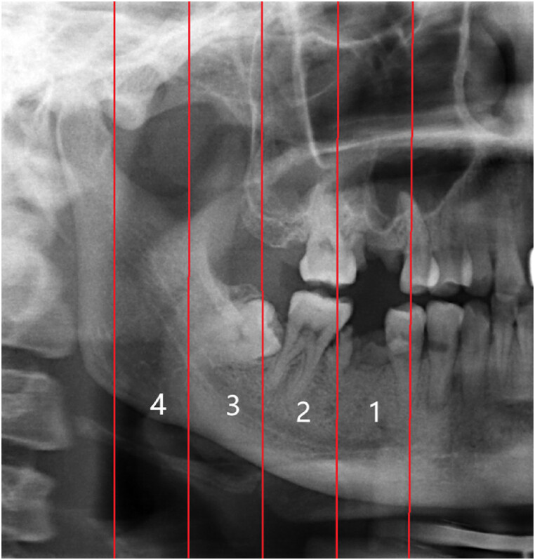
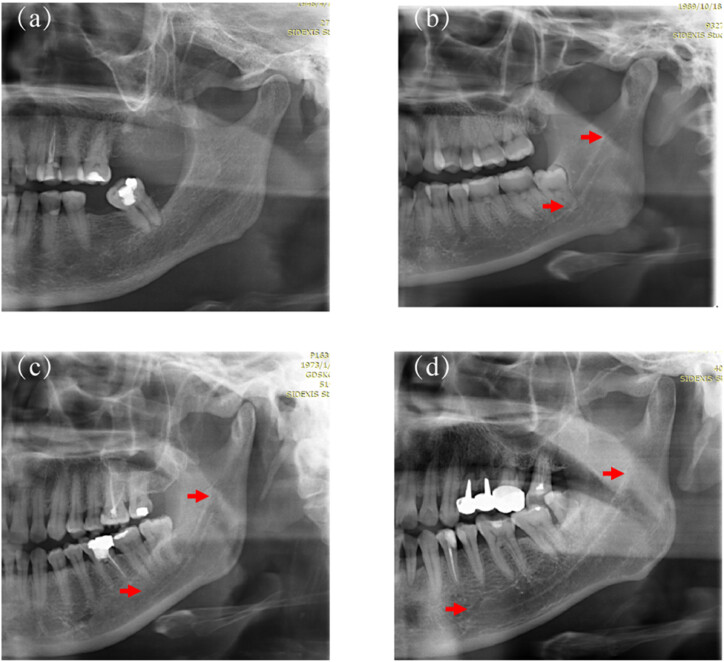
The testing dataset (n = 366) was divided into four groups based on image visibility to assess the performance of the CNN model and further compare it with the performance of two experienced radiologists. In a left/right PR image, IAC was bisected into four segments from the mental foramen to the mandibular foramen on average, defined as regions 1–4, respectively, as shown below. Based on the visibility of the four IAC regions, the validation dataset was grouped into: Group 1—the entire IAC is invisible (a); Group 2—less than half of IAC is clearly visible (b); Group 3—more than half of IAC is clearly visible (c); and Group 4—the entire IAC is clearly visible (d). One radiologist with more than ten years of experience manually grouped the PR images. The grouping was further reviewed and revised by a second radiologist with more than ten years of clinical experience in dentistry. A third radiologist was invited to settle the difference when the groupings of the two radiologists were not consistent.
Study Results
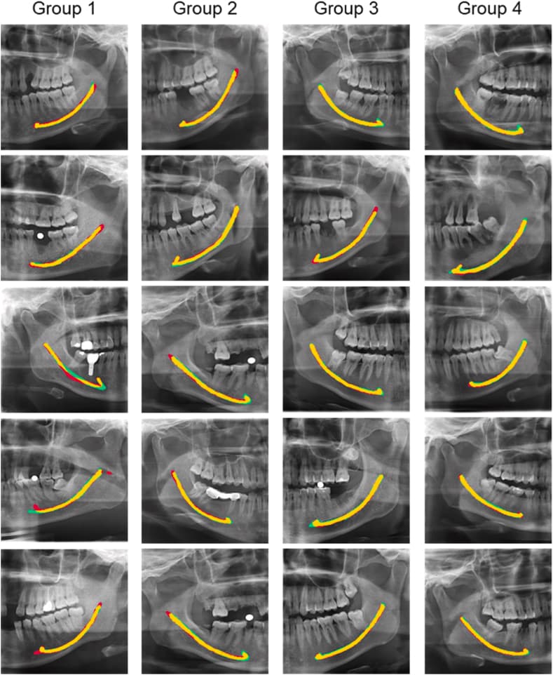
(Image on Left): The comparison between automated segmentation and manual annotation demonstrated that the IAC position was highly consistent between the two segmentation approaches, with a matching degree close to 85% These results demonstrated that CNN models have the potential to meet daily clinical needs. The edges of IAC predicted by the CNN model were more accurate than those predicted by manual delineation. In some special cases such as posterior maxillary area implants, posterior tooth root canal therapy, and even the postoperative review of the mandible, the location of IAC could be accurately distinguished by the CNN model.
(Image on Right) However, for some images with inaccurate predictions, especially for those in which the IAC boundary could not be discerned, there was a big difference between automated segmentation and manual annotation

Source of Images: Yang S, Li A, Li P, Yun Z, Lin G, Cheng J, Xu S, Qiu B. Automatic segmentation of inferior alveolar canal with ambiguity classification in panoramic images using deep learning. Heliyon. 2023 Feb 11;9(2):e13694.
List of Interesting Studies
Development of artificial intelligence model for supporting implant drilling protocol decision making., Takahiko Sakai , Hefei Li , Tatsuki Shimada , Suzune Kita , Maho Iida , Chunwoo Lee , Tamaki Nakano , Satoshi Yamaguchi , Satoshi Imazato , Journal of prosthodontic research. Volume: 67, Issue: 3, 2023
Purpose: This study aimed to develop an artificial intelligence (AI) model to support the determination of an appropriate implant drilling protocol using cone-beam computed tomography (CBCT) images.Methods:
Anonymized CBCT images were obtained from 60 patients. For each case, after implant placement, images of the bone regions at the implant site were extracted from 20 slices of CBCT images. Based on the actual drilling protocol, the images were classified into three categories: protocols A, B, and C. A total of 1,200 images were divided into training and validation datasets (n = 960, 80%) and a test dataset (n = 240, 20%). Another 240 images (80 images for each type) were extracted from the 60 cases as test data. An AI model based on LeNet-5 was developed using these data sets. The accuracy, sensitivity, precision, F-value, area under the curve (AUC) value, and receiver operating curve were calculated.Results The accuracy of the trained model is 93.8%. The sensitivity results for drilling protocols A, B, and C were 97.5%, 95.0%, and 85.0%, respectively, while those for protocols A, B, and C were 86.7%, 92.7%, and 100%, respectively, and the F values for protocols A, B, and C were 91.8%, 93.8%, and 91.9%, respectively. The AUC values for protocols A, B, and C are 98.6%, 98.6%, and 99.4%, respectively.
Conclusions:
The AI model established in this study was effective in predicting drilling protocols from CBCT images before surgery, suggesting the possibility of developing a decision-making support system to promote primary stability.
Convolutional neural network-based automated maxillary alveolar bone segmentation on cone-beam computed tomography images., Rocharles Cavalcante Fontenele , Maurício do Nascimento Gerhardt , Fernando Fortes Picoli , Adriaan Van Gerven , Stefanos Nomidis , Holger Willems , Deborah Queiroz Freitas , Reinhilde Jacobs , Clinical oral implants research. Volume: 34, Issue: 6, 2023
OBJECTIVES
To develop and assess the performance of a novel artificial intelligence (AI)-driven convolutional neural network (CNN)-based tool for automated three-dimensional (3D) maxillary alveolar bone segmentation on cone-beam computed tomography (CBCT) images.
MATERIALS AND METHODS:
A total of 141 CBCT scans were collected for performing training (n = 99), validation (n = 12), and testing (n = 30) of the CNN model for automated segmentation of the maxillary alveolar bone and its crestal contour. Following automated segmentation, the 3D models with under- or overestimated segmentations were refined by an expert for generating a refined-AI (R-AI) segmentation. The overall performance of CNN model was assessed. Also, 30% of the testing sample was randomly selected and manually segmented to compare the accuracy of AI and manual segmentation. Additionally, the time required to generate a 3D model was recorded in seconds (s).
RESULTS
The accuracy metrics of automated segmentation showed an excellent range of values for all accuracy metrics. However, the manual method (95% HD: 0.20 ± 0.05 mm; IoU: 95% ± 3.0; DSC: 97% ± 2.0) showed slightly better performance than the AI segmentation (95% HD: 0.27 ± 0.03 mm; IoU: 92% ± 1.0; DSC: 96% ± 1.0). There was a statistically significant difference of the time-consumed among the segmentation methods (p < .001). The AI-driven segmentation (51.5 ± 10.9 s) was 116 times faster than the manual segmentation (5973.3 ± 623.6 s). The R-AI method showed intermediate time-consumed (1666.7 ± 588.5 s).
CONCLUSION
Although the manual segmentation showed slightly better performance, the novel CNN-based tool also provided a highly accurate segmentation of the maxillary alveolar bone and its crestal contour consuming 116 times less than the manual approach.
Developing an Artificial Intelligence Solution to Autosegment the Edentulous Mandibular Bone for Implant Planning., Mohammad Adel Moufti , Nuha Trabulsi , Marah Ghousheh , Tala Fattal , Ali Ashira , Sebelan Danishvar , European journal of dentistry. Volume: 17, Issue: 4, 2023
OBJECTIVE: Dental implants are considered the optimum solution to replace missing teeth and restore the mouth's function and aesthetics. Surgical planning of the implant position is critical to avoid damage to vital anatomical structures; however, the manual measurement of the edentulous (toothless) bone on cone beam computed tomography (CBCT) images is time-consuming and is subject to human error. An automated process has the potential to reduce human errors and save time and costs. This study developed an artificial intelligence (AI) solution to identify and delineate edentulous alveolar bone on CBCT images before implant placement.MATERIALS AND METHODS
After obtaining the ethical approval, CBCT images were extracted from the database of the University Dental Hospital Sharjah based on predefined selection criteria. Manual segmentation of the edentulous span was done by three operators using ITK-SNAP software. A supervised machine learning approach was undertaken to develop a segmentation model on a “U-Net” convolutional neural network (CNN) in the Medical Open Network for Artificial Intelligence (MONAI) framework. Out of the 43 labeled cases, 33 were utilized to train the model, and 10 were used for testing the model’s performance.
STATISTICAL ANALYSIS
The degree of 3D spatial overlap between the segmentation made by human investigators and the model’s segmentation was measured by the dice similarity coefficient (DSC).
RESULTS
The sample consisted mainly of lower molars and premolars. DSC yielded an average value of 0.89 for training and 0.78 for testing. Unilateral edentulous areas, comprising 75% of the sample, resulted in a better DSC (0.91) than bilateral cases (0.73).
CONCLUSION
Segmentation of the edentulous spans on CBCT images was successfully conducted by machine learning with good accuracy compared to manual segmentation. Unlike traditional AI object detection models that identify objects present in the image, this model identifies missing objects. Finally, challenges in data collection and labeling are discussed, together with an outlook at the prospective stages of a larger project for a complete AI solution for automated implant planning.
Automated segmentation of the mandibular canal and its anterior loop by deep learning., Nicolly Oliveira-Santos , Reinhilde Jacobs , Fernando Fortes Picoli , Pierre Lahoud , Liselot Niclaes , Francisco Carlos Groppo , Scientific reports. Volume: 13, Issue: 1, 2023
Accurate mandibular canal (MC) detection is crucial to avoid nerve injury during surgical procedures. Moreover, the anatomic complexity of the interforaminal region requires a precise delineation of anatomical variations such as the anterior loop (AL). Therefore, CBCT-based presurgical planning is recommended, even though anatomical variations and lack of MC cortication make canal delineation challenging. To overcome these limitations, artificial intelligence (AI) may aid presurgical MC delineation. In the present study, we aim to train and validate an AI-driven tool capable of performing accurate segmentation of the MC even in the presence of anatomical variation such as AL. Results achieved high accuracy metrics, with 0.997 of global accuracy for both MC with and without AL. The anterior and middle sections of the MC, where most surgical interventions are performed, presented the most accurate segmentation compared to the posterior section. The AI-driven tool provided accurate segmentation of the mandibular canal, even in the presence of anatomical variation such as an anterior loop. Thus, the presently validated dedicated AI tool may aid clinicians in automating the segmentation of neurovascular canals and their anatomical variations. It may significantly contribute to presurgical planning for dental implant placement, especially in the interforaminal region. Read MoreAutomatic segmentation of inferior alveolar canal with ambiguity classification in panoramic images using deep learning., Shuo Yang , An Li , Ping Li , Zhaoqiang Yun , Guoye Lin , Jun Cheng , Shulan Xu , Bingjiang Qiu , Heliyon. Volume: 9, Issue: 2, 2023
BACKGROUND Manual segmentation of the inferior alveolar canal (IAC) in panoramic images requires considerable time and labor even for dental experts having extensive experience. The objective of this study was to evaluate the performance of automatic segmentation of IAC with ambiguity classification in panoramic images using a deep learning method.
METHODS Among 1366 panoramic images, 1000 were selected as the training dataset and the remaining 336 were assigned to the testing dataset. The radiologists divided the testing dataset into four groups according to the quality of the visible segments of IAC. The segmentation time, dice similarity coefficient (DSC), precision, and recall rate were calculated to evaluate the efficiency and segmentation performance of deep learning-based automatic segmentation.
RESULTS Automatic segmentation achieved a DSC of 85.7% (95% confidence interval [CI] 75.4%-90.3%), precision of 84.1% (95% CI 78.4%-89.3%), and recall of 87.7% (95% CI 77.7%-93.4%). Compared with manual annotation (5.9s per image), automatic segmentation significantly increased the efficiency of IAC segmentation (33 ms per image). The DSC and precision values of group 4 (most visible) were significantly better than those of group 1 (least visible). The recall values of groups 3 and 4 were significantly better than those of group 1.
CONCLUSIONS The deep learning-based method achieved high performance for IAC segmentation in panoramic images under different visibilities and was positively correlated with IAC image clarity.
Read More
A unique artificial intelligence-based tool for automated CBCT segmentation of mandibular incisive canal., Thanatchaporn Jindanil , Luiz Eduardo Marinho-Vieira , Sergio Lins de-Azevedo-Vaz , Reinhilde Jacobs , Dento maxillo facial radiology. Volume: 52, Issue: 8, 2023
OBJECTIVES To develop and validate a novel artificial intelligence (AI) tool for automated segmentation of mandibular incisive canal on cone beam computed tomography (CBCT) scans.METHODS After ethical approval, a data set of 200 CBCT scans were selected and categorized into training (160), validation (20), and test (20) sets. CBCT scans were imported into Virtual Patient Creator and ground truth for training and validation were manually segmented by three oral radiologists in multiplanar reconstructions. Intra- and interobserver analysis for human segmentation variability was performed on 20% of the data set. Segmentations were imported into Mimics for standardization. Resulting files were imported to 3-Matic for analysis using surface- and voxel-based methods. Evaluation metrics involved time efficiency, analysis metrics including Dice Similarity Coefficient (DSC), Intersection over Union (IoU), Root mean square error (RMSE), precision, recall, accuracy, and consistency. These values were calculated considering AI-based segmentation and refined-AI segmentation compared to manual segmentation.
RESULTS Average time for AI-based segmentation, refined-AI segmentation and manual segmentation was 00:10, 08:09, and 47:18 (284-fold time reduction). AI-based segmentation showed mean values of DSC 0.873, IoU 0.775, RMSE 0.256 mm, precision 0.837 and recall 0.890 while refined-AI segmentation provided DSC 0.876, IoU 0.781, RMSE 0.267 mm, precision 0. 852 and recall 0.902 with the accuracy of 0.998 for both methods. The consistency was one for AI-based segmentation and 0.910 for manual segmentation.
:CONCLUSIONS An innovative AI-tool for automated segmentation of mandibular incisive canal on CBCT scans was proofed to be accurate, time efficient, and highly consistent, serving pre-surgical planning.
Read More
Intra-oral scan segmentation using deep learning., Shankeeth Vinayahalingam , Steven Kempers , Julian Schoep , Tzu-Ming Harry Hsu , David Anssari Moin , Bram van Ginneken , Tabea Flügge , Marcel Hanisch , Tong Xi , BMC oral health. Volume: 23, Issue: 1, 2023
OBJECTIVE
Intra-oral scans and gypsum cast scans (OS) are widely used in orthodontics, prosthetics, implantology, and orthognathic surgery to plan patient-specific treatments, which require teeth segmentations with high accuracy and resolution. Manual teeth segmentation, the gold standard up until now, is time-consuming, tedious, and observer-dependent. This study aims to develop an automated teeth segmentation and labeling system using deep learning.
MATERIAL AND METHODS
As a reference, 1750 OS were manually segmented and labeled. A deep-learning approach based on PointCNN and 3D U-net in combination with a rule-based heuristic algorithm and a combinatorial search algorithm was trained and validated on 1400 OS. Subsequently, the trained algorithm was applied to a test set consisting of 350 OS. The intersection over union (IoU), as a measure of accuracy, was calculated to quantify the degree of similarity between the annotated ground truth and the model predictions.
RESULTS
The model achieved accurate teeth segmentations with a mean IoU score of 0.915. The FDI labels of the teeth were predicted with a mean accuracy of 0.894. The optical inspection showed excellent position agreements between the automatically and manually segmented teeth components. Minor flaws were mostly seen at the edges.
CONCLUSION The proposed method forms a promising foundation for time-effective and observer-independent teeth segmentation and labeling on intra-oral scans.
CLINICAL SIGNIFICANCE : Deep learning may assist clinicians in virtual treatment planning in orthodontics, prosthetics, implantology, and orthognathic surgery. The impact of using such models in clinical practice should be explored. ::![]()
Read More
Neural network system for analyzing statistical factors of patients for predicting the survival of dental implants., Pavel Alekseevich Lyakhov , Alexander Alexandrovich Dolgalev , Ulyana Alekseevna Lyakhova , Alexandr Alexandrovich Muraev , Kirill Evgenievich Zolotayev , Dmitry Yurievich Semerikov , Frontiers in neuroinformatics. Volume: 16, Issue: , 2022
Implants are now the standard method of replacing missing or damaged teeth. Despite the improving technologies for the manufacture of implants and the introduction of new protocols for diagnosing, planning, and performing implant placement operations, the percentage of complications in the early postoperative period remains quite high. In this regard, there is a need to develop new methods for preliminary assessment of the patient's condition to predict the success of single implant survival. The intensive development of artificial intelligence technologies and the increase in the amount of digital information that is available for analysis make it relevant to develop systems based on neural networks for auxiliary diagnostics and forecasting. Systems based on artificial intelligence in the field of dental implantology can become one of the methods for forming a second opinion based on mathematical decision making and forecasting. The actual clinical evaluation of a particular case and further treatment are carried out by the dentist, and AI-based systems can become an integral part of additional diagnostics. The article proposes an artificial intelligence system for analyzing various patient statistics to predict the success of single implant survival. As the topology of the neural network, the most optimal linear neural network architectures were developed. The one-hot encoding method was used as a preprocessing method for statistical data. The novelty of the proposed system lies in the developed optimal neural network architecture designed to recognize the collected and digitized database of various patient factors based on the description of the case histories. The accuracy of recognition of statistical factors of patients for predicting the success of single implants in the proposed system was 94.48%. The proposed neural network system makes it possible to achieve higher recognition accuracy than similar neural network prediction systems due to the analysis of a large number of statistical factors of patients. The use of the proposed system based on artificial intelligence will allow the implantologist to pay attention to the insignificant factors affecting the quality of the installation and the further survival of the implant, and reduce the percentage of complications at all stages of treatment. However, the developed system is not a medical device and cannot independently diagnose patients. At this point, the neural network system for analyzing the statistical factors of patients can predict a positive or negative outcome of a single dental implant operation and cannot be used as a full-fledged tool for supporting medical decision-making. Read MoreArtificial intelligence and augmented reality for guided implant surgery planning: A proof of concept., Francesco Guido Mangano , Oleg Admakin , Henriette Lerner , Carlo Mangano , Journal of dentistry. Volume: 133, Issue: , 2023
PURPOSE
To present a novel protocol for authentic three-dimensional (3D) planning of dental implants, using artificial intelligence (AI) and augmented reality (AR).
METHODS
The novel protocol consists of (1) 3D data acquisition, with an intraoral scanner (IOS) and cone-beam computed tomography (CBCT); (2) application of AI for CBCT segmentation to obtain standard tessellation language (STL) models and automatic alignment with IOS models; (3) loading of selected STL models within the AR system and surgical planning with holograms; (4) surgical guide design with open-source computer-assisted-design (CAD) software; and (5) surgery on the patient.
RESULTS
This novel protocol is effective and time-efficient when used for planning simple cases of static guided implant surgery in the partially edentulous patient. The clinician can plan the implants in an authentic 3D environment, without using any radiological guided surgery software. The precision of implant placement looks clinically acceptable, with minor deviations.
CONCLUSIONS
AI and AR technologies can be successfully used in guided implant surgery for authentic 3D planning that may replace conventional software. However, further clinical studies are needed to validate this protocol.
STATEMENT OF CLINICAL RELEVANCE
The combined use of AI and AR may change the perspectives of modern guided implant surgery for authentic 3D planning that may replace conventional software. ::![]()
Read More
Influence of bone density on the accuracy of artificial intelligence-guided implant surgery: An in vitro study., Zhicong Chen , Yun Liu , Xin Xie , Feilong Deng , The Journal of prosthetic dentistry. Volume: 131, Issue: 2, 2024
STATEMENT OF PROBLEM
Artificial intelligence (AI) has been found to be applicable in medical tests and diagnostics. However, studies on the application of AI technology in oral implantology are lacking. In addition, whether bone density affects the accuracy of guided implant surgery has not been determined.
PURPOSE
The purpose of this in vitro study was to determine the clinical reliability of an AI-assisted implant planning software program with an in vitro model. An additional goal was to determine the effect of bone density on the accuracy of static computer-assisted implant surgery (CAIS).
MATERIAL AND METHODS
Ten participants with missing mandibular left first molars were selected for analysis, and surgical fully guided templates were designed by using an AI implant planning software program. Jaw models were produced in 3 filling rate groups (group L: 25%; group M: 40%; group H: 55%, higher filling rate with representatives of the denser simulated bone density) by 3-dimensional (3D) printing. The preoperative and postoperative positions of the implants were compared by measuring the value of deviation through oral scanning. The mean 3D shoulder and apical and angular deviations were calculated for each group. The data were analyzed using 1-way ANOVA (α=.05 corrected for multiple testing by using Bonferroni-Holm adjustment).
RESULTS
The mean ±standard deviation 3D shoulder and apical and angular deviations were 0.80 ±0.32 mm, 1.43 ±0.47 mm, and 3.68 ±1.30 degrees. These values were lower than the clinical safety distance of the fully guided implant template. A significantly lower mean 3D apical deviation (1.12 ±0.33 mm, P=.023) and angular deviation (2.81 ±1.11 degrees, P=.018) were observed in group L than in group H (1.68 ±0.37 mm, 4.32 ±0.99 degrees). However, no significant differences were found among the 3 groups in 3D deviation at the shoulder (P>.05).
CONCLUSIONS
AI implant planning software program could design the ideal implant position through self-learning. The accuracy of the AI-assisted designed implant template in this study indicated its clinical reliability. Higher bone density led to increased implant deviations.