This Week in Dental Implants, following a new study on Soft-Tissue Augmentation in the Aesthetic Zone, we wanted to review the concepts around Soft-Tissue Augmentation around implants, and including presentation of a few case studies that demonstrate some of the popular techniques.
Note: For additional information about management of defects around implants, please see an earlier case: Effectively Addressing Defects around Implants with a Cortical Bone Sheet
Soft Tissue Management Review: What are the key benefits?
Although, there is still some debate around the impact of various soft tissue deficiencies, soft‑tissue management is generally considered important for the long‑term success of dental implants because it influences:
| Aspect | Why it matters |
|---|---|
| Implant survival and stability | A healthy peri‑implant soft tissue seal protects the underlying bone and helps maintain osseointegration. |
| Esthetic outcome | Adequate width, thickness and keratinized tissue provide a more natural‑looking emergence profile and reduce recession, especially in the anterior (esthetic) zone. |
| Biological health | Soft‑tissue augmentation improves the formation of a stable barrier (biological width) that resists bacterial invasion; insufficient tissue can lead to peri‑implant mucositis or peri‑implantitis. |
| Maintenance and hygiene | Keratinized mucosa facilitates patient cleaning, reduces discomfort and helps prevent plaque‑related problems. |
| Predictability of surgical results | Proper timing of soft‑tissue grafting (pre‑, post‑surgical, before or after loading) allows clinicians to tailor treatment to patient needs and to avoid complications such as dehiscence or recession. |
| Healing dynamics | Soft‑tissue wound healing around implants requires 6–8 weeks for barrier formation; managing the tissue promotes proper maturation and reduces scar‑like peri‑implant mucosa. |
| Patient quality of life | Successful soft‑tissue management improves comfort, function and patients’ confidence in their prosthetic result. |
Latest Research: Soft Tissue Augmentation Improves Peri-Implant Soft Tissue Thickness, But Not Buccal Bone Remodeling
One of the key factors influencing long-term implant success is peri-implant soft tissue thickness (STT). STT refers to the horizontal thickness of the mucosa surrounding a dental implant, regardless of whether the tissue is keratinized. Research suggests that a minimum thickness of approximately 2 mm is desirable. From an esthetic perspective, tissue at least 2 mm thick helps mask the underlying implant components and reduces the risk of gray shine-through. Thicker soft tissue has also been associated with improved papilla fill, less gingival recession over time, and higher patient satisfaction. Biologically, increasing soft tissue thickness through grafting has been linked to better peri-implant health and reduced marginal bone loss. 2
A recent randomized clinical trial 4 compared connective tissue grafts (CTG), a volume-stable collagen matrix (VCMX), and no soft tissue augmentation during implant placement in the esthetic zone. Both CTG and VCMX significantly increased peri-implant soft tissue thickness compared with no grafting, with VCMX showing slightly greater tissue gains. However, neither approach prevented the normal remodeling and loss of buccal bone that occurred after implant placement. Patients reported similar outcomes across all groups, although CTG resulted in slightly greater postoperative discomfort.
Clinical takeaway: Soft tissue augmentation is effective for increasing peri-implant soft tissue thickness, which may improve esthetic and biological outcomes. However, based on this study, increasing STT alone does not prevent buccal bone remodeling following implant placement.
Case Photos: Review of Main Soft Tissue Management Techniques
All photos below are from Sun TC, Chang TK. Soft tissue management around dental implant in esthetic zone - the current concepts and novel techniques.
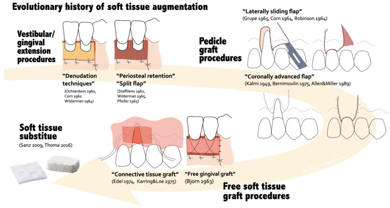
The evolutionary history of soft tissue augmentation around natural dentition, the techniques has been further applied to soft tissue management around dental implants.
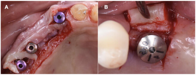
Flap techniques for soft tissue management around dental implants. (A) The Palacci flap technique (B) The roll flap technique.
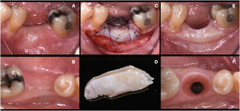
Free gingival graft surgical procedure to increase the width of keratinized mucosa around dental implants (A and B) Clinical presentation at baseline (C) Stabilization of free gingival graft at recipient bed (D) Free gingival graft harvest from palate (E and F) Twelve-month outcomes, notice the significant increased width of soft tissue and keratinized tissue.
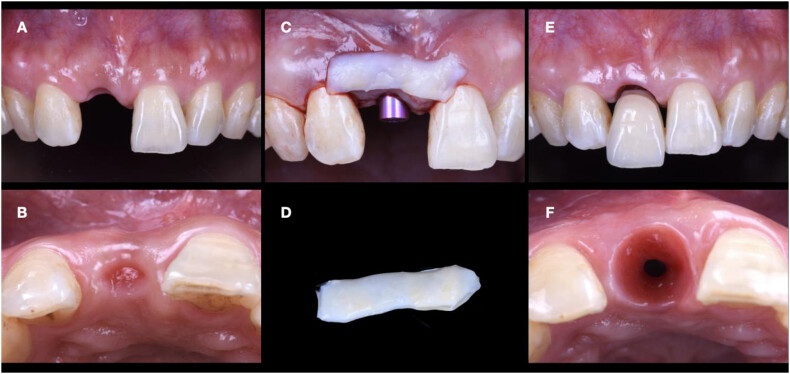
Connective tissue graft surgical procedure around dental implant to increase the peri-implant soft tissue thickness (A and B) Clinical presentation at baseline (C and D) Connective tissue graft harvest from palate ready to be inserted into recipient bed (E and F) Twelve-month outcomes, notice the significant increased width of peri-implant soft tissue and improved esthetic.
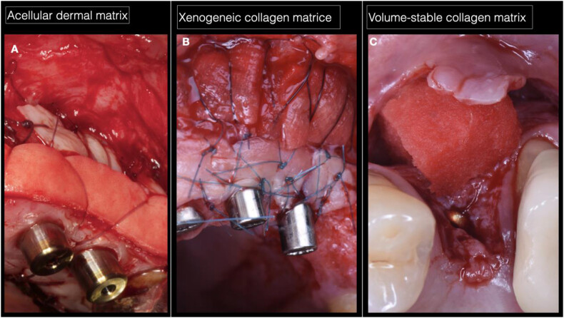
Application of the soft tissue substitutes for soft tissue management around dental implants (A) Combination use of acellular dermal matrix and free gingival graft to increase keratinized mucosa for extensive soft tissue deficiency (B) Combination use of xenogeneic collagen matrix and free gingival graft to increase keratinized mucosa for extensive soft tissue deficiency (C) Application of volume-stable collagen matrix to increase peri-implant soft tissue thickness.
Case: Prevention of soft tissue complications with implant placement
All case photos below are from: Management and prevention of soft tissue complications in implant dentistry.
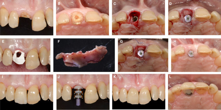
Clinical case of soft tissue management simultaneous to implant placement and guided bone regeneration. A‐B, Patient’s initial situation. C, Immediate implant placement after extraction of tooth 11. D, Guided bone regeneration performed buccal to implant 11. E, Collagen membrane adapted to seal the socket. F, Sub‐epithelial connective tissue graft. G, Partial thickness pouch buccal to the implant with autogenous graft. H, Post‐operative healing after 3 months. I, Implant‐supported provisional restoration. J, Conventional open tray impression of implant 11. K‐L, Implant‐supported restoration on 11 after one year follow‐up.
Case: Prevention of soft tissue complications during implant‐healing phase
All case photos below are from: Management and prevention of soft tissue complications in implant dentistry.
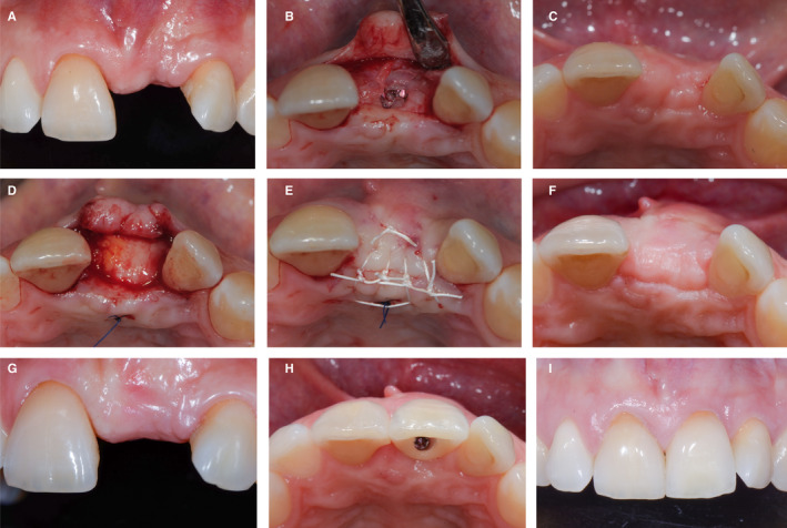
Clinical case of soft tissue management within the implant healing phase, after implant placement and before abutment connection. A, Patient’s initial situation with missing tooth 21. B, Implant placement without augmentation on 21. C, Post‐operative healing after one month. D, Placement of a sub‐epithelial connective tissue graft buccal and occlusal to the implant. E, Primary wound closure with sutures. F‐G, Post‐operative healing after 2 months. H, Implant‐supported restoration on 21. I, Clinical situation at three year follow‐up
Case: Prevention of soft tissue complications during abutment connection
All case photos below are from: Management and prevention of soft tissue complications in implant dentistry.
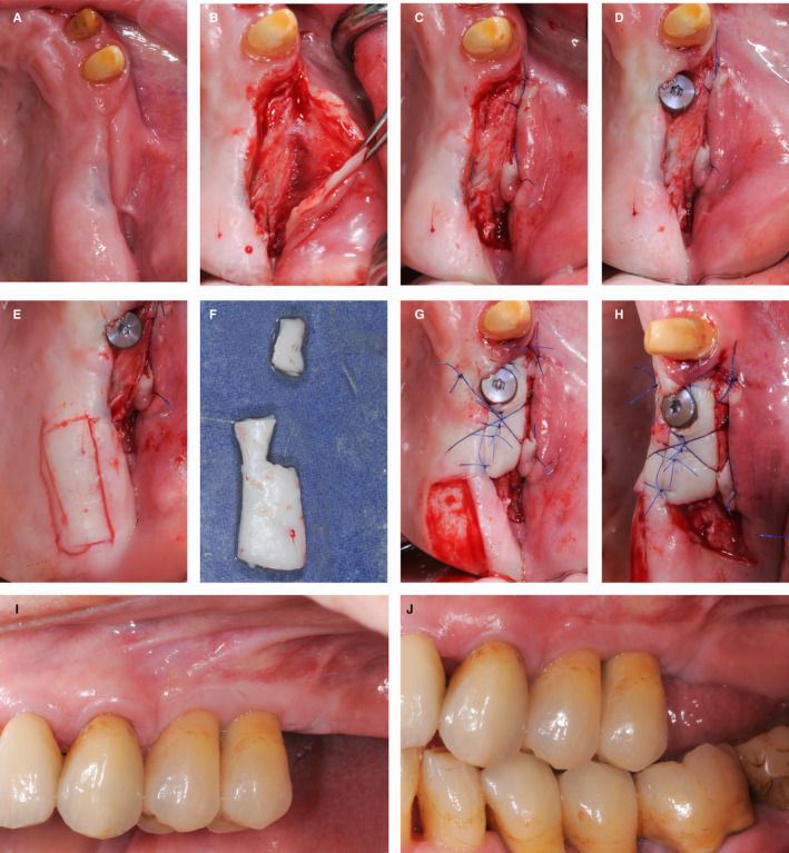
Clinical case of soft tissue management at abutment connection. A, Patient’s initial situation. B, Partial thickness incision buccal to placed implant 24. C, Apical suturing of the flap. D, Healing abutment placed on implant 24. E, Free gingival graft from the palate. F, Trimming of the free gingival graft. G‐H, Stabilization of the free gingival graft with sutures. I, Post‐operative healing after 4 weeks. J, Clinical situation after 2 years of follow‐up
References
- Sun TC, Chang TK. Soft tissue management around dental implant in esthetic zone - the current concepts and novel techniques. J Dent Sci. 2024 Jul;19(3):1348-1358.
- Moussa H, Nasri W, Gargouri R, Bouslema A. Management of Soft Tissue Defects Around Single Implants: A Systematic Review of the Literature. Clin Exp Dent Res. 2024 Dec;10 (Note: This is an excellent article that review the various types of soft tissue deficiencies).
- Thoma DS, Gil A, Hämmerle CHF, Jung RE. Management and prevention of soft tissue complications in implant dentistry. Periodontol 2000. 2022 Feb;88(1):116-129.
- Papantonatou L, Batas L, Delantoni A, Anagnostou E, Vouros I. Soft-Tissue Augmentation During Implant Placement in the Aesthetic Zone: Clinical, Radiographic, Histological and Patient-Reported Outcomes From a Randomised Controlled Trial Using Connective Tissue Graft and a Volume-Stable Collagen Matrix. J Clin Periodontol. 2026 Jul 28.