This Week in Dental Implants, we want to highlight 2 new studies that deal with drilling techniques in implant dentistry. Drilling technique can significantly affect osseointegration by influencing the integrity of the osteotomy site, the bone-to-implant contact, and the mechanical properties of the surrounding bone, such as density and quality. Therefore, selecting an appropriate drilling method is essential for optimizing the success of dental implants.
Given the importance of drilling technique, we were intrigued by these new studies:
- Effects of Asymmetric Oscillatory Drilling on Bone Densification, Heat Generation, and Implant Stability., which concluded that: “Oscillatory movement during implant site preparation enhanced bone densification, improved primary stability, and maintained safe thermal levels. These findings suggest that oscillatory movement could serve as a promising alternative to conventional drilling techniques, particularly in low-density bone.”
- Optimizing Osseodensification Drilling for Dental Implant Placement: An In Vitro Study., which provided the following clinical implication: “For Type IV alveolar bone, OD at 1500 rpm and 0.04 mm/z is recommended for improving the primary stability of dental implants. Sufficient irrigation is crucial in both CD and OD for circumventing the thermal damage to bone.”
With regards to Osseodensification, for which there is wide body of research (see some references provided below), the method involves the use of specially designed burs that compact and densify bone along the osteotomy walls during implant site preparation. Unlike traditional drilling methods that remove bone, osseodensification preserves and compacts the bone. This compaction increases the density of the surrounding bone, which can potentially enhance the primary stability of the implant.
Case Photos
Case 1: Osseodensification Drilling for Dental Implant Placement:
Case photos provided by: Tao X, Yang J, Ma T, Chen M, An Q, Yu D. Optimizing Osseodensification Drilling for Dental Implant Placement: An In Vitro Study. Clin Exp Dent Res. 2025 Jun;
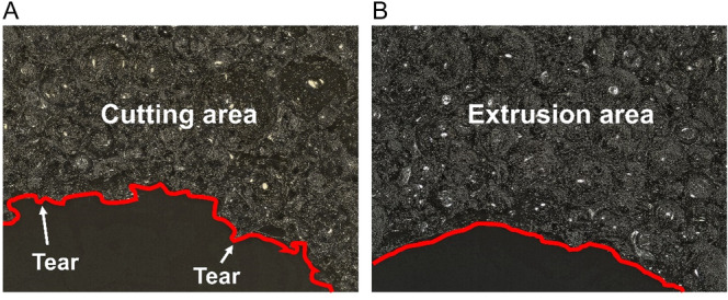
Characteristics of prepared implant sites. (A) implant sites prepared via BLT drills; (B) implant sites prepared via Densah drills (osseodensification). The entrance characteristics of implant sites were assessed using a material‐type upright laser confocal microscope. The entrance characteristics were varied when using different drills to prepare the implant sites under the same condition (𝑁 = 300 r/min, 𝐹𝑧 = 0.08 mm/z, depth = 10 mm, without irrigation). In the CD group, burrs, tears, and potholes were observed at the prepared sites. However, only a few burrs appeared in the OD group.
Case 2: Osseodensification Drilling in the Narrow Alveolar Ridge:
Case photos provided by: Shanmugam M, Valiathan M, Balaji A, Jeyaraj Samuel AF, Kannan R, Varthan V. Conventional Versus Osseodensification Drilling in the Narrow Alveolar Ridge: A Prospective Randomized Controlled Trial. Cureus. 2024 Mar 26;16(3):e56963. doi: 10.7759/cureus.56963.
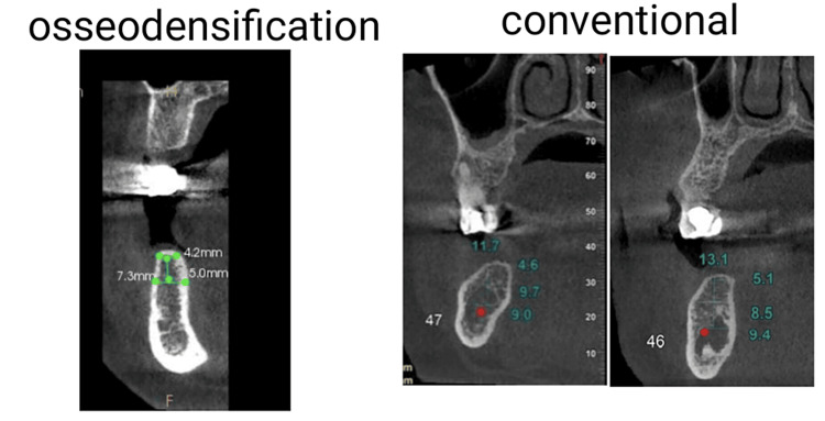
Figure 1. Preoperative cone-beam computed tomography showing narrow alveolar ridge in both the osseodensification protocol and conventional protocol. The pre-surgical assessment was conducted following standard protocols, and preoperative 3D CBCT was performed to evaluate buccolingual width, bone quality, and height of the alveolar ridge. Buccolingual width of less than 6 mm was considered in the study
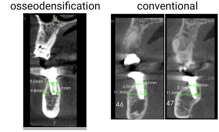
Figure 2: Six-month postoperative cone-beam computed tomography to evaluate alveolar ridge expansion. During the six months following CBCT, there was no difference in crestal width between the conventional and osseodensification drilling groups, even though the latter approach induced increased diameter in the narrow ridge during osteotomy. However, when measured 5 mm from the apex to the crest, there was a substantial difference in the width in the osseodensification group, with the conventional group exhibiting more variability. Both sets of data demonstrated a statistically significant improvement in radiographic bone density. Both groups had a statistically significant increase in implant stability over time, as determined by ISQ values.
References
1. Effects of Asymmetric Oscillatory Drilling on Bone Densification, Heat Generation, and Implant Stability., Henrique Tadeu do Rego Ferreira , Emmanuel João Nogueira Leal da Silva , Bruno Salles Sotto Maior , Plinio Mendes Senna , The International journal of oral & maxillofacial implants. Volume: 0, Issue: 0, 2025
PURPOSE
Achieving optimal implant site preparation is crucial for successful osseointegration, particularly in low-density bone. Excessive heat generation during drilling can compromise bone vitality, leading to impaired healing, delayed osseointegration or implant failure. This study evaluates the effects of an oscillatory drilling technique on bone densification, heat generation, and implant stability.
MATERIALS AND METHODS
An in vitro study was conducted using pig rib segments to simulate low-density bone. Drilling protocols were performed usin three techniques: clockwise cutting (CW), counterclockwise densifying (CC), and oscillatory movement (OM) at 60° clockwise/240° counterclockwise. Drilling was performed with automated handpiece handling under controlled conditions. Thermal changes were recorded using a laser thermometer and infrared imaging, and bone densification was assessed using micro-CT analysis. The final insertion torque (IT) was measured to determine the primary stability and removal torque was recorded to access the stability loss for each implant.
RESULTS
The CC and OM groups demonstrated significantly higher bone volume, intersection surface, and reduced trabecular separation compared to the CW group (p < 0.05). Although the CC group exhibited the highest temperature increase, all final temperatures remained below the critical threshold of 47°C. Infrared imaging showed overall less heat retained within the body of the drill in CW group compared to CC and OM. OM drills exhibited lower heat generation than the CC group with temperature peak approximately 5 mm above the drill tip. The OM and CC groups showed significantly higher IT than the CW group (p < 0.05). Implant removal revealed a stability reduction of 27.8±10.2%, 24.8±13.6%, and 20.2±8.2% for CW, CC and OM groups, respectively (p > 0.05).
CONCLUSIONS
Oscillatory movement during implant site preparation enhanced bone densification, improved primary stability, and maintained safe thermal levels. These findings suggest that oscillatory movement could serve as a promising alternative to conventional drilling techniques, particularly in low-density bone. Further studies are needed to explore its clinical applicability and long-term outcomes. Read More
2. Optimizing Osseodensification Drilling for Dental Implant Placement: An In Vitro Study., Xingru Tao , Jie Yang , Tai Ma , Ming Chen , Qinglong An , Dedong Yu , Clinical and experimental dental research. Volume: 11, Issue: 3, 2025
OBJECTIVES
This study aims to optimize the parameters of osseodensification drilling (OD) to improve the primary stability of dental implants in low-density bone.
MATERIAL AND METHODS
Polyurethane foam blocks (PFB) of 0.160 g/cm, were used to simulate low-density bone (Type IV bone). Two drills, i.e., bone level tapered drills and Densah drills, were used in conventional drilling (CD) and OD, respectively. Drilling was performed on a DMU machine, varying spin speed, feed per tooth, and irrigation with or without 4°C saline. Thrust force, temperature, and entrance characteristics of implant sites in CD and OD were compared. The primary stability of implants was assessed via insertion torque (IT), removal torque (RT), and implant stability quotient (ISQ).
RESULTS
OD demonstrated higher thrust force than CD with maximum values at 300 rpm (2.99 ± 0.22 N vs. 2.77 ± 0.17 N, p < 0.01). Temperature elevation was lower in OD than that in CD under irrigation (3.35°C vs. 4.67°C). Despite comparable ISQ values (CD: 46.71 ± 8.56 vs OD: 47.08 ± 5.95, p = 0.86), OD achieved higher IT (11.73 ± 0.45 N·m vs. 7.77 ± 0.21 N·m, p < 0.001) and higher RT (9.28 ± 0.45 N·m vs. 6.65 ± 0.19 N·m, p < 0.001). Morphological analysis revealed fewer defects (tears/potholes) in OD than CD.
CONCLUSIONS
OD drills may avoid iatrogenic damage of the morphology of implant sites and enhance primary stability in Type IV bone at 1500 rpm and 0.04 mm/z with irrigation to prevent thermal damage. OD also outweights CD in increased bone density, thrust force, and torque.
CLINICAL IMPLICATIONS
For Type IV alveolar bone, OD at 1500 rpm and 0.04 mm/z is recommended for improving he primary stability of dental implants. Sufficient irrigation is crucial in both CD and OD for circumventing the thermal damage to bone. Read More
3. Comparative evaluation of osseodensification drilling versus conventional drilling technique on dental implant stability: A systematic review., Saurav Banerjee , Dolanchanpa Dasgupta , Nikita Parasrampuria , Dipankar Pal , Udey Vir Gandhi , Journal of Indian Prosthodontic Society. Volume: 24, Issue: 3, 2024
AIM
The present systematic review compares the stability, crestal bone levels and efficacy of osseodensification (OD) drilling techniques for dental implant placement to traditional drilling methods.
SETTINGS AND DESIGN
The Cochrane online library, PubMed, Scopus, and other well-known online resources are used in the research. Using a systematic review design, the current study examines published qualitative studies with an emphasis on analysis.
MATERIALS AND METHODS
Using precise keywords, a thorough search of pertinent databases was carried out in accordance with PRISMA standards. Studies testing dental implant stability, crestal bone levels and clinical results using both OD and traditional procedures were covered by the inclusion criteria.
STATISTICAL ANALYSIS USED
The risk of bias and quality of included studies was assessed using the Newcastle-Ottawa Scale for observational studies and the Cochrane Risk of Bias tool for randomized controlled trials.
RESULTS
A total of 170 patients and 334 implants from Egypt, India, and Brazil were included in eight papers that made up the systematic review. In several clinical situations, osseodensification outperformed standard drilling in terms of implant durability, bone development, and torque data. Statistical analysis presented the lowest risks, while blinded outcome assessment, allocation concealment, random sequence generation, incomplete outcome data and experimental technique revealed higher risks. Bias assessment found various risks across different components.
CONCLUSION
The thorough examination of eight papers demonstrates that osseodensification is a technique with great promise in the field of dental implants. It exhibits superior torque values, bone development, and stability when compared to traditional drilling. The overall results highlight the potential of osseodensification to improve clinical outcomes and advance the science of dental implantology, even in the face of variances in bias concerns. Read More
4. Implant Stability of Osseodensification Drilling Versus Conventional Surgical Technique: A Systematic Review., João Gaspar , Luís Proença , João Botelho , Vanessa Machado , Leandro Chambrone , Rodrigo Neiva , José João Mendes , The International journal of oral & maxillofacial implants. Volume: 36, Issue: 6, 2021 Nov-Dec
PURPOSE
This systematic review aimed to appraise the available evidence on the clinical characteristics produced by osseodensification drilling compared with the conventional drilling technique.
MATERIALS AND METHODS
Five databases (PubMed, Google Scholar, LILACS, EMBASE, and CENTRAL) were searched up to July 2020. Randomized clinical trials (RCTs) and nonrandomized studies of interventions (NRSIs) that compared osseodensification drilling with conventional drilling in humans were included. Random-effects meta-analyses of standardized mean difference (MD) with 95% confidence intervals (CI) and risk ratio were performed.
RESULTS
Three NRSIs fulfilled the inclusion criteria, and all were scored as low risk of bias. Meta-analysis showed that the osseodensification drilling technique presented higher average implant stability quotient (ISQ) scores at baseline (MD: 13.1, 95% CI: 10.0 to 16.1, P < .0001) than conventional drilling, with complete homogeneity (I, = 0.0%). Furthermore, osseodensification drilling presented higher average ISQ scores at follow-up (MD: 5.99, 95% CI: 1.3 to 10.6, P < .0001) than conventional drilling, with high homogeneity (I, = 73.0%).
CONCLUSION
This systematic review showed that osseodensification presented consistently higher ISQ at baseline and at 4 to 6 months after implant placement compared with conventional drilling. However, these results should be carefully interpreted since only three studies were selected in this meta-analysis. In the future, RCTs will be necessary to confirm the consistency of these results. Read More
5. Conventional Versus Osseodensification Drilling in the Narrow Alveolar Ridge: A Prospective Randomized Controlled Trial., Mohanasatheesh Shanmugam , Mohan Valiathan , Anitha Balaji , Angelin Fiona Jeyaraj Samuel , Rudra Kannan , Vishnu Varthan , Cureus. Volume: 16, Issue: 3, 2024
Background Conventionally, undersized osteotomies were used to increase initial bone-to-implant contact to achieve primary stability in implantology. This is particularly evident in regions with low bone density. The potential for severe bone compression and ischemia poses a challenge to secondary stability. Instead, lateral bone compaction is caused by the idea of osseodensification. Research on the potential benefits of this method for narrow ridges is lacking. This study aimed to determine if the osseodensification drilling technique affects primary stability and how much the alveolar ridge expands following implant site preparation. Methodology A total of 30 participants aged 20 to 80 years were included in this randomized controlled clinical investigation. Each participant was randomly assigned to one of the following two groups: one that received standard drill preparation, and another that received osseodensification drill preparation. Implant stability using implant stability quotient values, crest width, apical width (5 mm from crest), and bone density were assessed both before and after six months using cone-beam computed tomography. Results Osseodensification impacted the width at the apex (5 mm from the crest) and radiographic bone density, adding to the quality, but did not affect implant stability and crestal width after osseointegration. The mean difference in conventional and osseodensification groups was 0.46 and 0.68 mm, respectively, concerning the crestal width. Moreover, the mean difference was 0.74 and 0.58 mm for conventional and osseodensification groups, respectively, concerning the width at the apex (5 mm from the crest). Conclusions This study demonstrates that the osseodensification process increased both the radiographic bone density and the width at the apex, demonstrating that osseodensification drilling techniques allow for the placement of implants with larger diameters in narrow alveolar ridges. Read More6. Biomechanical and histomorphometric analysis of endosteal implants placed by using the osseodensification technique in animal models: A systematic review and meta-analysis., Amit M Gaikwad , Amruta A Joshi , Jyoti B Nadgere , The Journal of prosthetic dentistry. Volume: 127, Issue: 1, 2022
STATEMENT OF PROBLEM
Osseodensification, a counterclockwise drilling technique for the placement of endosseous implants is a popular clinical technique. However, the effect of the osseodensification technique on primary implant stability, bone-implant contact, and bone area frequency occupancy is unclear.
PURPOSE
The purpose of this systematic review and meta-analysis was to investigate the biomechanical and histomorphometric outcomes of endosteal implants placed by using the osseodensification technique in animal models.
MATERIAL AND METHODS
An electronic search through Medline/PubMed, Lilacs, and Science Direct databases, and an additional manual search of the reference list of included articles was conducted by using specific keywords and Medical Subject Headings (MeSH) terms for articles in the English language and published up to April 31, 2020. Only animal studies comparing the biomechanical and histomorphometric outcomes of endosteal implants placed by using the osseodensification and conventional drilling protocol were included. The SYstematic Review Center for Laboratory animal Experimentation (SYRCLE) tool was used to determine the risk of bias assessment, and the quality of included studies was assessed by using Animal Research: Reporting in Vivo Experiments (ARRIVE) guidelines.
RESULTS
Nine studies were included. The results of the meta-analysis showed that the pooled weighted mean difference of the insertion torque value for the primary implant stability of endosseous dental implants placed by using the osseodensification technique was 2.270 (95% confidence interval [CI]=1.147 to 3.393; P<.001), the weighted mean difference of the percentage of bone-implant contact at 3 weeks was 0.487 (95% CI=0.220 to 0.754; P=.114), the weighted mean difference of the percentage of bone-implant contact at 6 weeks was 0.565 (95% CI=0.219 to 0.911; P=.448), the weighted mean difference of the percentage of bone area frequency occupancy at 3 weeks was 0.679 (95% CI=0.265 to 1.093; P=.073), and the weighted mean difference of the percentage of bone area frequency occupancy at 6 weeks was 0.391 (95% CI=-0.204 to 0.986; P=.027).
CONCLUSIONS
Limited data from animal studies suggest that the primary implant stability, bone-implant contact, and bone area frequency occupancy significantly improved for the endosteal implants placed by using the osseodensification technique compared with conventional drilling protocol. However, additional laboratory and clinical studies are recommended to provide stronger evidence. Read More