This Week in Dental Implants we are following up on a new study on Marginal Bone Level (MBL) Changes following Implant placement and highlighting some additional cases surrounding the MBL issue.
Introduction
Marginal bone loss (MBL) around dental implants is a significant concern in implant dentistry as it can affect the long-term success and stability of the implants. There are many factors that can potentially influence MBL and these encompass both surgical reasons (insertion torque, improper Implant position etc.) and prosthetic techniques (type of connection, presence and location of implant/abutment microgap, abutment height, remaining cement etc.), as well as other issues such as bone density, gingival biotype, smoking, diabetes, and bruxism. Properly understanding the causes of MBL and managing these risk factors through careful planning is therefore essential to help minimize marginal bone loss and improve the longevity of dental implants.
New Study: Correlation of Implant Location to Marginal Bone Level Changes in Single-Unit Restorations
A new study) 1 of 226 patients with single-unit dental implants in molar, premolar, and anterior provided really interesting results. It suggested that the probability of bone loss in premolar implants is over 2 times higher than in molar implants and that the probability of bone loss in anterior implants is over 3 times higher than in molar implants. The study concluded that:
The findings of this study indicate that MBL changes can be evaluated over a range of Emergence Angle (EA) values. It can be stated that as the duration of prosthetic delivery time increases following the surgical placement of dental implants, the risk of marginal bone loss also increases. 1
Case Study: Marginal bone loss in immediate dental implants
Case photos below provided by Rehberger Bescós F, Salgado Peralvo ÁO, Chamorro Petronacci CM, Chele D, Camacho Alonso F, Peñarrocha Oltra D, Lado Baleato Ó, Pérez Sayáns M. [Marginal bone loss and associated factors in immediate dental implants: a retrospective clinical study.](Marginal bone loss and associated factors in immediate dental implants: a retrospective clinical study - PMC)
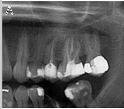
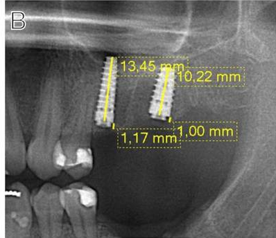
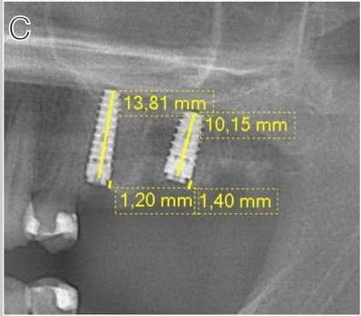
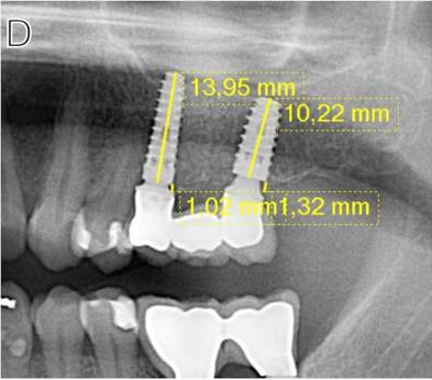
Measurements for the calculation of the MBL performed: A Preoperative evaluation; B immediate postoperative period; C 2 months after placement of the implants; D 36 month follow-up
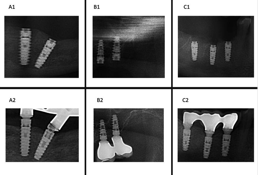
A BL example in IDI #33 (A1: March 2019- A2: September 2023); B BR example in IDI #26 (B1: November 2019- B2: January 2020); C BO example in IDI #46 (C1: January 2019-C2 January 2020)
This study concluded:
The present study supports the clinical efficacy of immediate implant placement protocol with high survival rates and acceptable MBL. Regarding the latter, the insertion of implants bone level about 3 mm infracrestal should be considered to ensure a subcrestal implant platform position during bone remodeling during the first months after implantation. The insertion of immediate implants in the jaw compared to the maxilla, the abutment height and rotational abutments demonstrated a positive impact over the MBL.
Case Study: Effect of vertical implant position on marginal bone loss
Case photos below provided by: Hedayatipanah M, Arasteh HK, Shokri A, Alafchi B, Baghdadi LS. Effect of vertical implant position on marginal bone loss: a randomized clinical trial.
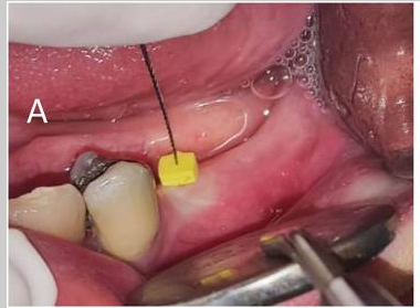
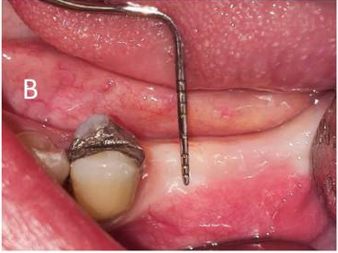
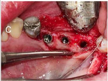

(A) Soft tissue thickness measurement (B) Keratinized gingiva measurement (C) Implant placement (D) Three months after implant placement and healing abutment closure
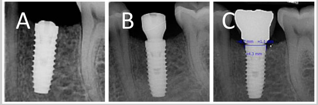
Crestal implant: (A) baseline (B) three months after placement (C) three months after loading
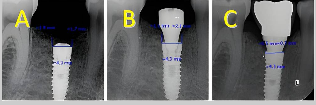
Subcrestal implant: (A) baseline (B) three months after placement (C) three months after loading
This study concluded:
The relationship between implant’s vertical position, soft tissue thickness and marginal bone loss is complex. While this research suggests that subcrestal placement and thinner tissues potentially lead to greater bone loss, more research is needed to fully understand the individual and combined effects of these factors. The time factor also significantly affects the amount of marginal bone loss, so the loss in the third month after implant placement is more than the loss in the third month after implant placement (six months after implant placement).
Case Study: Assessing marginal bone loss around mandibular posterior dental implants
Case photo below provided by: Sharma K, Aggarwal S, Gupta K, Madan R, Tariq S, Narula IS, Gupta S. Three-Dimensional Evaluation of Alveolar Bone Levels Around Dental Implants and Natural Teeth: A Prospective Study.
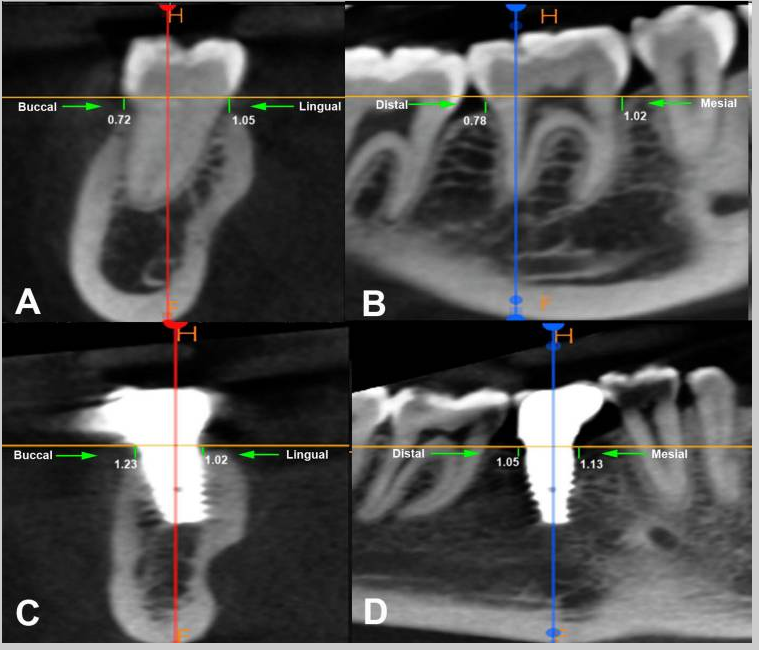
Evaluation of the marginal bone loss: (A) Buccal and lingual aspect of natural tooth, (B) mesial and distal aspect of natural tooth, (C) buccal and lingual aspect of dental implant, (D) mesial and distal aspect of dental implant.
This study concluded that:
Based on the findings of the present study, it was concluded that the mean marginal bone loss and probing depth( PD) were higher on the implant side than on the natural contralateral tooth during the 18-month follow-up period, and it was the highest on the distal side. Bone loss and PD were higher at the 18-month follow-up than at the six-month follow-up. The duration between the first- and second-stage implant surgery and PD were two significant predictors of bone loss at the mesial and distal sides of the implant. Age and sex were not significantly correlated with bone loss in any of the regions.
References
- Ayyildiz BG, Ayyildiz H, Tözüm TF. Correlation of Implant Location to Marginal Bone Level Changes in Single-Unit Restorations: A Retrospective Study. Clin Implant Dent Relat Res. 2025 Oct;27(5):e70092.
- Rehberger Bescós F, Salgado Peralvo ÁO, Chamorro Petronacci CM, Chele D, Camacho Alonso F, Peñarrocha Oltra D, Lado Baleato Ó, Pérez Sayáns M. Marginal bone loss and associated factors in immediate dental implants: a retrospective clinical study. Int J Implant Dent. 2025 Mar 26;11(1):25.
- Hedayatipanah M, Arasteh HK, Shokri A, Alafchi B, Baghdadi LS. Effect of vertical implant position on marginal bone loss: a randomized clinical trial. BMC Oral Health. 2024 Jun 24;24(1):727.
- Sharma K, Aggarwal S, Gupta K, Madan R, Tariq S, Narula IS, Gupta S. Three-Dimensional Evaluation of Alveolar Bone Levels Around Dental Implants and Natural Teeth: A Prospective Study. Cureus. 2024 Oct 9;16(10):e71129.