Introduction: Membrane Fixation Overview
A fundamental principle of guided bone regeneration (GBR) is maintaining a stable, protected space where new bone can develop. Membrane fixation is the step of securing a barrier membrane to the surrounding bone so that it stays immobile over the graft, as well as to prevent displacement of the graft . Various techniques have been suggested to achieve adequate fixation, such as screws, pins, or special suture procedures, e.g. periosteal vertical mattress suture.
Based on current studies, membrane fixation is not universally required. For example, in well‑contained defects fixation may not change overall ridge volume, 1 but in many other graft situations (non‑contained defects, larger defects, or when maximal bone formation is desired) fixation can enhance membrane stability, increase osteogenic factor expression, and improve new‑bone formation. 2 The benefit of fixation may also be membrane‑type dependent, with some collagen membranes showing coronal width gain with fixation, while others showing no effects. 3 Additionally, some kinds of porcine collagen membranes (like our OsseoSeal membrane), have amazing adaptability when wet and adhere to the site, thereby obviating the need for any type of fixation technique. Amnion-chorion membranes also exhibit unmatched adhesion and therefore wouldn’t really benefit from fixation.
But, when fixation is indicated how do the different methods actually compare quantitatively? That is the subject of the this Weeks highlighted study.
Recent Study: Comparing No-Fixation (NF), Periosteal sutures (PS), or Titanium fixation pins
In this study, researchers evaluated three methods of stabilizing collagen membranes during horizontal guided bone regeneration in non-contained ridge defects using fresh human cadaver models. The membrane was either left unsecured, stabilized with periosteal sutures, or fixed with titanium pins.
Titanium fixation pins provided the greatest stability, virtually eliminating graft displacement during flap closure and preserving over 80% of the grafted volume. Periosteal sutures also significantly improved stability compared with no fixation, although they were less effective than pins. Sites without membrane fixation experienced the greatest graft movement, volume loss, and ridge collapse.
The study concluded:
Even after passive flap release and tension-free closure, membrane fixation, particularly with titanium pins, provides superior resistance to graft displacement in non-contained defects, while unfixed membranes allow substantial graft loss and are therefore not recommended in non-contained defects. 4
Featured Case: Combining Periosteal Suture with Four Pins
In this case below, 5 a autogenous graft covered with absorbable membranes were fixed by both periosteal diagonal mattress sutures combined with four corner pins.
The researchers concluded:
This case highlights that PDMS combined with four corner pins is feasible to maintain the space and stabilize the graft and membranes in severe continuous horizontal bone defect…Titanium pins and sutures can be combined effectively and flexibly. Compared with the sausage technique using pins only, multiple pins are required, which is costly, and it is difficult to operate the pins on the lingual or palatal side, and sometimes it is also difficult to avoid hazardous sites and damage to adjacent roots during insertion. Reducing the number of titanium pins can reduce the risk of complications and the difficulty of surgery. This combined technique is flexible and is very useful in clinical practice.
Case photos below provided by: Wang LH, Ruan Y, Zhao WY, Chen JP, Yang F. Modified membrane fixation technique in a severe continuous horizontal bone defect: A case report.
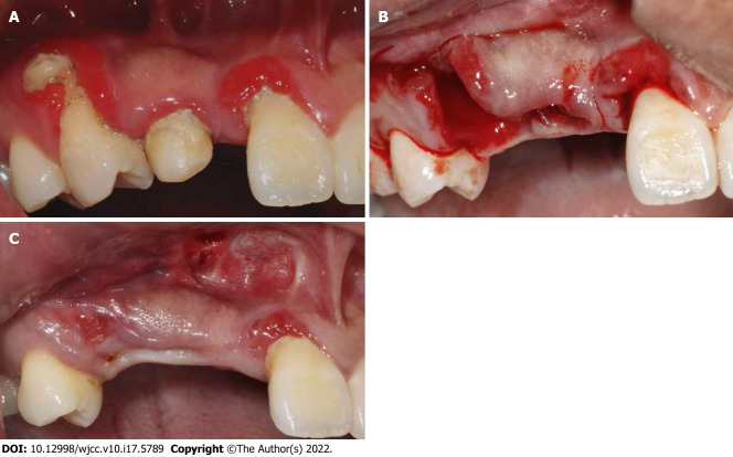
Oral examination of the patient at initial visit, after tooth extraction and before bone grafting. A: Initial situation of the edentulous site in maxillary anterior; B: Extraction of teeth 53 and 14; C: Three weeks after teeth extraction and before bone augmentation surgery.
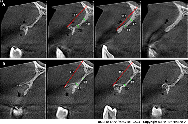
Bone volume at the defect area before bone grafting. A: Cone beam computed tomography (CBCT) before bone augmentation surgery 5 and 10 mm below the alveolar crest at the site of missing tooth 12; B: CBCT before bone augmentation surgery 5 and 10 mm below the alveolar crest at the site of missing tooth 14.
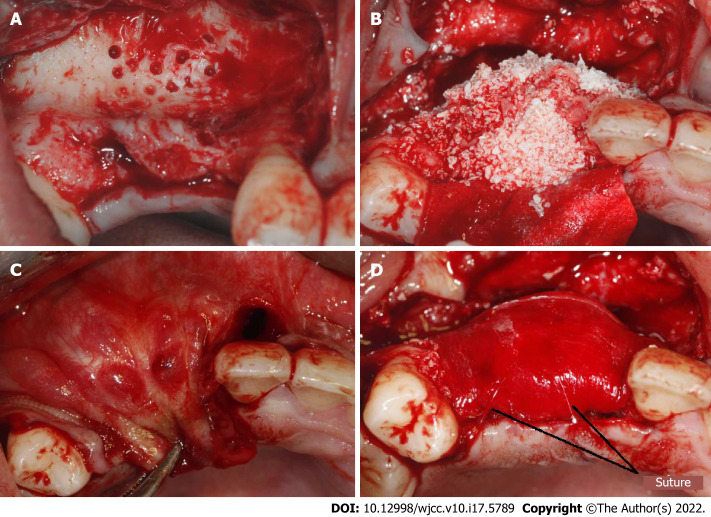
Procedure of bone grafting. A: Decortication holes were prepared at the recipient area; B: Bone graft material was placed; C: Make sure the soft tissue could be primary closed without tension; D: The resorbable membranes and bone grafts were fixed using periosteal diagonal mattress sutures and four corner pins.
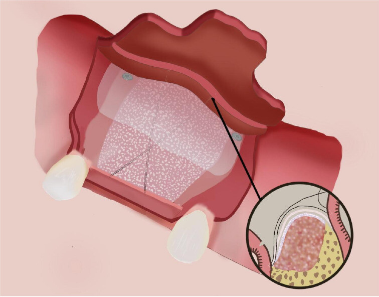
Fixation of the membrane using periosteal diagonal mattress suture and four corner pins.
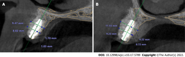
Cone beam computed tomography superimposed images before bone grafting and after implantation. A: At site of implant 12. The yellow line represented the alveolar ridge before bone grafting. The bone width was increased from 1.83 to 8.83 mm at a point 5 mm below the crest, and from 1.70 to 9.47 mm at a point 10 mm below the crest; B: At site of implant 14. The yellow line represented the alveolar ridge before bone grafting. The bone width was increased from 0.72 to 9.23 mm at a point 5 mm below the crest, and from 4.22 to 11.55 mm at a point 10 mm below the crest.
References
- Park JY, Song YW, Ko KA, Strauss FJ, Thoma DS, Lee JS. Effect of collagen membrane fixation on ridge volume stability and new bone formation following guided bone regeneration. J Clin Periodontol. 2022 Jul;49(7):684-693.
- An YZ, Strauss FJ, Park JY, Shen YQ, Thoma DS, Lee JS. Membrane fixation enhances guided bone regeneration in standardized calvarial defects: A pre-clinical study. J Clin Periodontol. 2022 Feb;49(2):177-187.
- Park JY, Song YW, Ko KA, Strauss FJ, Thoma DS, Lee JS. Effect of collagen membrane fixation on ridge volume stability and new bone formation following guided bone regeneration. J Clin Periodontol. 2022 Jul;49(7):684-693.
- Saleh MHA, Calatrava J, Zambrana N, Parma-Benfenati L, Sabri H, Urban I, Wang HL. Collagen Membrane Fixation Technique Affects Its Space Maintenance Capacity After Flap Closure: An Ex Vivo Analysis. Clin Oral Implants Res. 2026 Jun 12.
- Wang LH, Ruan Y, Zhao WY, Chen JP, Yang F. Modified membrane fixation technique in a severe continuous horizontal bone defect: A case report. World J Clin Cases. 2022 Jun 16;10(17):5789-5797.