This Week in Dental Implants we are highlighting a study on the accuracy of cone-beam computed tomography (CBCT) for assessing distances between implants and the mandibular canal.
Background: CBCT and Nerve canals in the Mandibular Region.
Proper assessment of implant proximity to the mandibular canal is a key factor in preventing nerve-related complications during implant placement. According to one study:
The safe zone recommended for implant surgery is 4 mm anterior and 8 mm inferior to the mental foramen, and 10 mm above the inferior margin of mandible. The chin bone should be harvested at least 10 mm below the tooth apices along with a limited depth of 4 mm. 1
Unlike traditional two-dimensional imaging, CBCT provides a three-dimensional view of dental anatomy, allowing for more accurate assessment of the mandibular canal and improved implant planning. This can potentially help reduce the risk of implant placement errors and injury to adjacent structures.
New Study: Accuracy of CBCT for assessing distances between implant and mandibular canal?
However, despite the growing popularity of CBCT, a new study casts some doubt on its ultimate accuracy for small distances.2. It concluded that:
CBCT cannot accurately identify implant-canal distances ≤ 0.4 mm, which may directly affect clinical risk assessment. Even for distances ≥ 0.5 mm, CBCT underestimates the true distance by 0.2-0.3 mm. These findings provide practical guidance for setting safe margins in implant planning and postoperative evaluation.
Featured Case: Evaluation of mandibular canal visibility on cross-sectional cone-beam CT images
In this study, 360 total CBCT cross-sectional images were examined from Indiana University School of Dentistry Imaging Facility. 3
Case images below from Comparative evaluation of mandibular canal visibility on cross-sectional cone-beam CT images: a retrospective study.
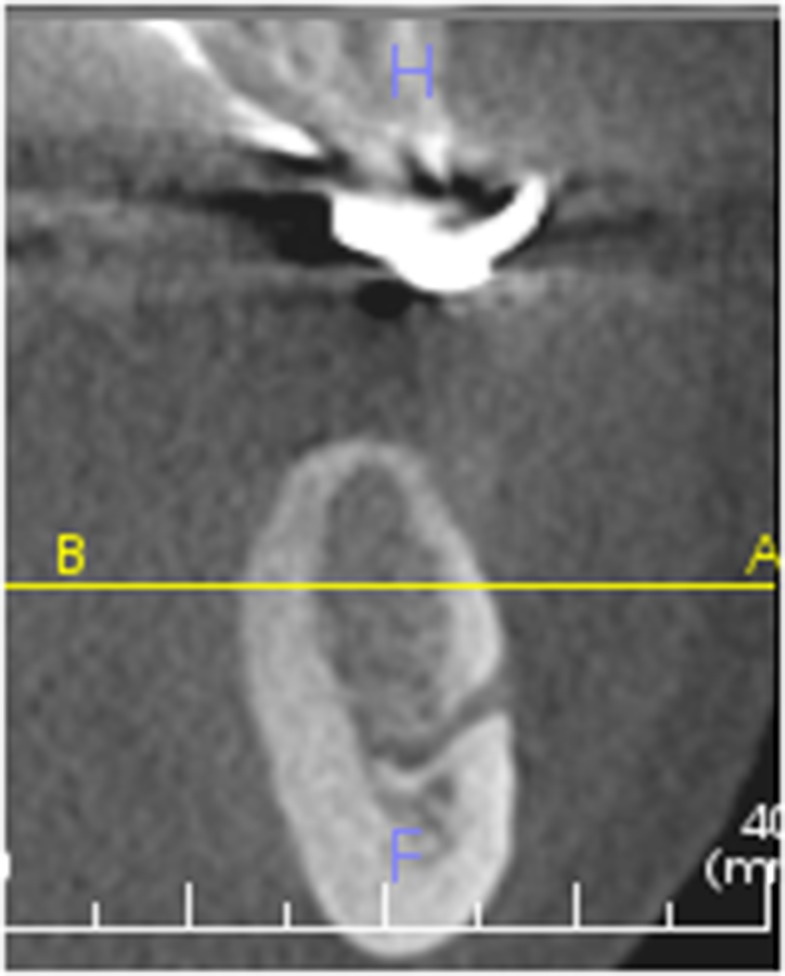
Mental foramen visible at PM1 41 × 52 mm (96 × 96 dots per inch).
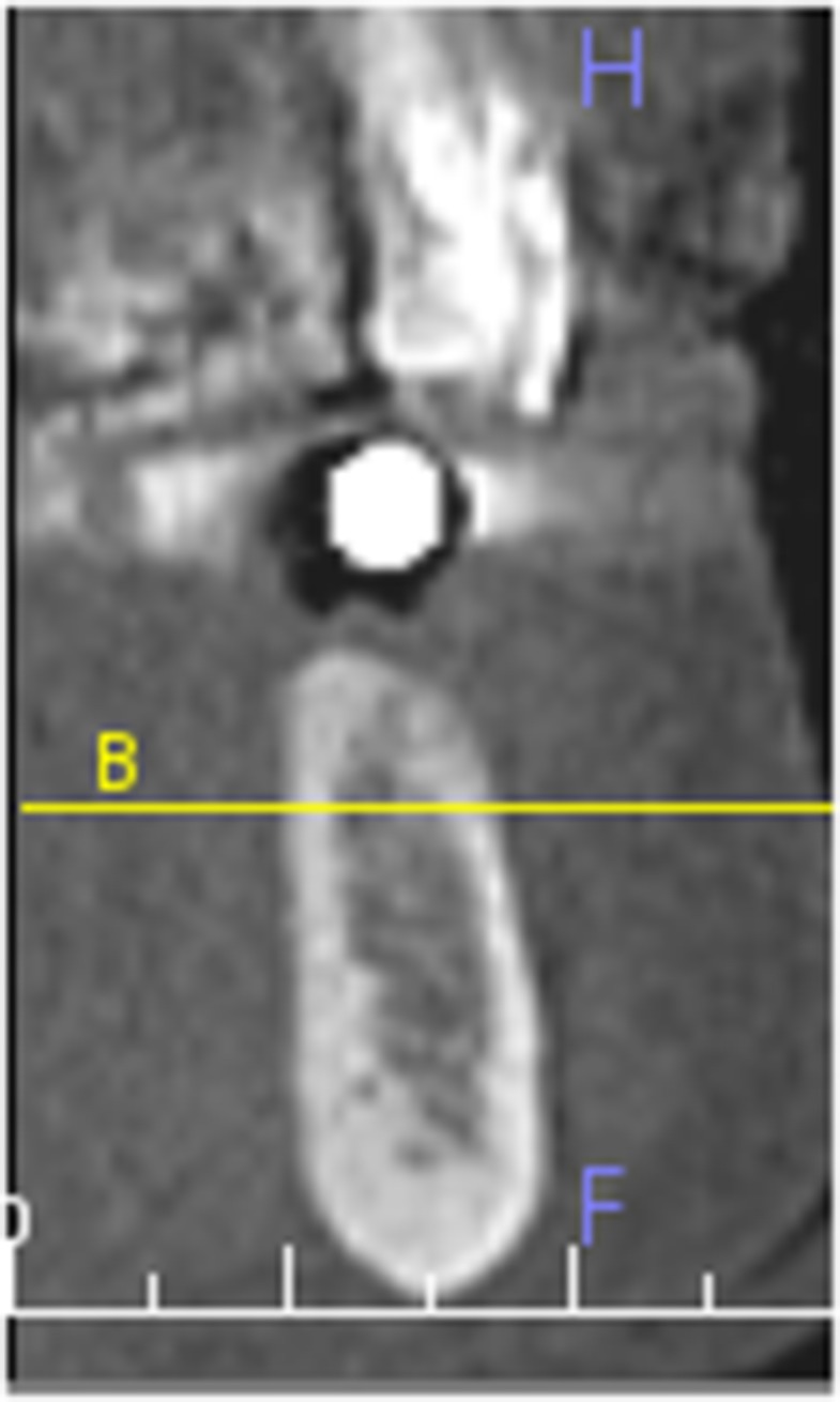
Mandibular canal visible at M2 36 × 50 mm (96 × 96 dots per inch).
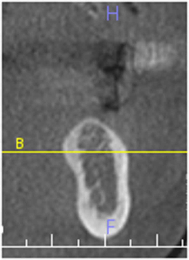
Mandibular canal not distinguished from other anatomical structures at PM1 30 × 51 mm (96 × 96 dots per inch).
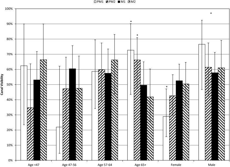
Canal visibility (%) by age and location, with 95% confidence interval. Significant visibility differences were found: female < male, PM1, age 47–56 < PM1 age 65+, M2 age 65+ < PM1 age 65 + and PM2 age 65+, (p = 0.0180); female PM1 < female M1 and female M2. M1, first molar; M2, second molar; PM1, first premolar; PM2, second premolar.
This study concluded that: 3
It is important to visualize the MC in cross-sectional CBCT images for implant planning. Moreover, having this knowledge aids clinicians in making better evidence-based decisions with regards to prescribing imaging studies for patients and minimizing the radiation dose whenever possible while getting the essential information from the proper imaging modality. Identification of the MC in 56% of the CBCT cross-sectional images is similar to results reported in previous studies. Although CBCT imaging is a valuable asset in the assessment of sites for implant placement, it is unable to consistently provide visualization and identification of the MC in all instances, and careful evaluation of the implant site is necessary to avoid impingement or violation of vital structures. The conclusion of this retrospective study was that the MC was identified in just over half of the time in selected, single CBCT cross-sectional images. Age, gender and location had significant effects on the visibility of the MC. CBCT cross-sectional imaging is a valuable tool for identification of vital anatomic structures as part of treatment planning but is not without limitations for identification of all anatomic structures.
Featured Case: Investigating the characteristics of the mandibular canal in cone beam CT
This was a retrospective study of 112 CBCT images of Vietnamese patients aged 18 to 69 years, taken for clinical indications between 2018 and 2023. 4
> Case images below from: Investigating the characteristics of the mandibular canal in cone beam CT. J Orthod Sci.
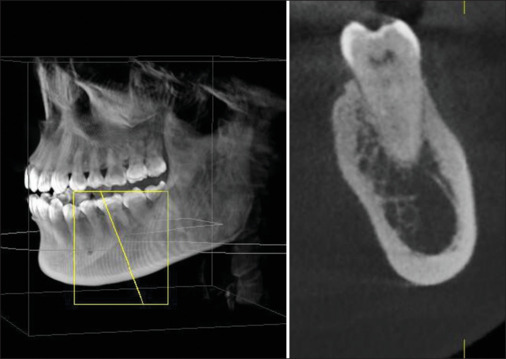
The sagittal plane has been appropriately rotated to pass through the apex and intersect perpendicular to the axis of the MC at the location of the mesial apex of the first molar
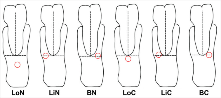
Guidelines for assessing the correlation of the MC. LiC - Lingual contact, BC - Buccal contact, LoC - Lower contact, LiN - lingual noncontact, BN - buccal noncontact and LoN - Lower noncontact
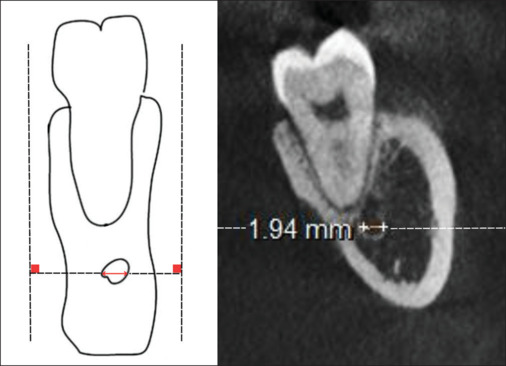
Corresponding diameter of the MC at the mesial apex of the first molar
This study concluded as follows:
The precise localization of the MC relative to the tooth apex and the diameter of the MC can vary in each person. CBCT provides cross-sectional images that can help surgeons with oral disease diagnosis and treatment processes. CBCT indications should be considered when establishing treatment planning to avoid damaging the IAN block in the MC. Otherwise, when CBCT conditions are not ideal, having enough knowledge about anatomical structures is necessary to realize the risk of bias during treatment.
Can AI Help with the accuracy of segmenting the mandibular canal?
Another interesting development in the accuracy of CBCT is in the ability of AI to improve the results. In this study 5, localization of the mandibular canals was performed for 104 randomly selected patients, and concluded that:
An AI-driven segmentation of the mandibular canal constitutes a time-efficient and reliable procedure for pre-operative implant planning.
Of course, it’s still early for AI applications in this area, but additional advancements will certainly help bridge the gap, and provide a more accurate assessment to avoid clinical errors.
References
- Yang XW, Zhang FF, Li YH, Wei B, Gong Y. Characteristics of intrabony nerve canals in mandibular interforaminal region by using cone-beam computed tomography and a recommendation of safe zone for implant and bone harvesting. Clin Implant Dent Relat Res. 2017 Jun;19(3):530-538.
- Yoshihara H, Honda E, Matsuka Y. Measurement accuracy of dental cone-beam computed tomography for assessing submillimeter distances between implant and mandibular canal: a phantom study. Int J Implant Dent. 2026 Jun 7.
- Miles MS, Parks ET, Eckert GJ, Blanchard SB. Comparative evaluation of mandibular canal visibility on cross-sectional cone-beam CT images: a retrospective study. Dentomaxillofac Radiol. 2016;45(2):20150296.
- Do TT, Le LN, Tan LT, Dang ATT, Huynh DNK, Truong MH, Nguyen LM. Investigating the characteristics of the mandibular canal in cone beam CT. J Orthod Sci. 2024 Nov 25;13:45.
- Ntovas P, Marchand L, Finkelman M, Revilla-León M, Att W. Accuracy of artificial intelligence-based segmentation of the mandibular canal in CBCT. Clin Oral Implants Res. 2024 Sep;35(9):1163-1171.