Dental implants in the esthetic zone have consistently demonstrated high survival rates, but achieving a successful outcome involves juggling many complex issues. This Week in Dental Implants we are highlighting new research, involving more than 14,000 implants, that provides a real-world look at survival in the anterior maxilla. Interestingly, the study reveals that the factors associated with failure can differ depending on when it occurs.
Implants in the Esthetic Zone: What are some of the Issues?
Placing implants in the esthetic zone is especially demanding due to several biological, surgical and prosthetic challenges. Specifically, some issues to consider are:
- Technique sensitivity: Immediate placement and restoration are technique‑sensitive; any minor aberration can produce major esthetic and functional problems, especially in younger patients who need long‑term stability.
- Hard and soft tissues preservation: After extraction the buccolingual bone width collapses, and the marginal bone and gingival levels must be maintained to keep natural contours and papillae.
- Primary stability: Low insertion torque may require a custom healing abutment, while high torque demands an immediate provisional to hold the buccal bone graft and preserve tissue shape.
- Accurate positioning: The implant’s vertical, sagittal and buccal placement must be ideal to support the emergence profile and avoid compromising the interproximal papilla.
- Managing limited bone volume: Narrow ridges often need bone‑manipulation techniques (ridge splitting, osseodensification, socket‑shield, guided bone regeneration, titanium mesh, etc.) that add complexity and lengthen treatment.
- Controlling implant design factors – Implant morphology (tapered vs. cylindrical) and the drill‑implant diameter discrepancy affect rotational stability, which is critical when the implant is placed immediately and provisionalized.
Survival of Implants in the Esthetic Zone: What does the prior research show?
Despite these challenges, dental implants placed in the esthetic zone generally demonstrate high survival rates. The best summary we found of the data was a 2024 meta-analysis, which concluded:
The overall failure rate was found to be 2% (95% CI; 0.00-0.03%). The percentage marginal bone loss was estimated to be 1% (95% CI; 0.00 - 0.02%) and the mean and proportion pink esthetic scores were approximately 11.75 (0.43%) with 2% mid-facial soft tissue recession and the mesial and distal papillary recession was 0.02% and 0.01%, respectively. Based on this systematic review and meta-analysis, the rate of dental implant failure for implant placement in the esthetic zone was minimal. 6
Real-World Survival in the Esthetic Zone: The Latest Study
In Real-World Implant Survival in the Esthetic Zone: Uncovering Timing-Specific Risk Patterns in a Large Cohort 1, Haim etc al. analyzed electronic health-record data from a nationwide dental network, examining 14,079 implants in 8,234 patients treated between 2014 and 2023. The study focused on implants placed in the anterior maxilla and followed patients for a mean of 6.0 ± 2.9 years. 1. During that period, 358 implants failed. That corresponds to an overall failure rate of 2.5%, remarkably similar to the approximately 2% failure rate reported in a 2024 systematic review. 6
But, what was unique about the Haim et al. study was that rather than treating every failure as the same event, they divided failures into different phases of treatment. Using a six-month cutoff, they distinguished early failures from late failures and then examined which factors were associated with each phase.
This produced some interesting data:
-
Of the 358 failures in the study, 43.6% occurred during the early phase.
-
Immediate restoration was associated with an increased risk of early failure. The reported hazard ratio was 1.91, meaning that within the statistical model used by the researchers, immediate restoration was associated with approximately 91% higher hazard of early failure compared with the reference group.
-
Smoking, however, showed a different pattern. It was associated with late failure, with a reported hazard ratio of 1.81.
-
The study also identified short implants, defined as *8 mm or less, as being associated with failure across the different phases. However, this finding needs to be interpreted cautiously because the short-implant subgroup was very small, containing only 54 implants.
The key insight from this study appears to be that, similar to previous research, implant placement in the esthetic zone demonstrates high long-term survival in general, but implant failure cannot necessarily be viewed as a single clinical event with a single set of risk factors. Instead, the factors associated with failure appear to differ depending on when the failure occurs. Immediate restoration was associated with early failure, while smoking was associated with late failure, and short implant length (≤8 mm) was associated with failure across both phases. So each of these risk factors needs to be considered clinically along the life of the implant.
We should note in closing that the 2026 study is retrospective, so it cannot establish that the identified factors directly caused implant failure. Associations observed in medical-record data can also be influenced by variables that are difficult to fully control. Nevertheless, the size of the cohort is significant. More than 14,000 anterior maxillary implants provide a much broader view of implant survival than many individual clinical studies or case series.
Cases: Surgical Techniques for Successful Placement of Implants in the Esthetic Zone
The cases below are from Enhancing Anterior Esthetic Zone Implant Placement Through Bone Manipulation Techniques: A Case Series. Indu Raj Harinee A Anjana S Raj Arvind K Uikey Femitha Syed et al.
Rehabilitation of maxillary anterior teeth is a challenging task especially when a patient presents with bone defects. Bone augmentation using autogenous bone grafts followed by implant placement is the gold standard treatment for such patients to achieve desirable esthetics. But more often than not, patients may not be comfortable with the delay until the bone maturation. Hence, a few viable alternatives were brought into the forum such as lateral bone expansion using thread formers of increasing diameters, osseodensification using densah burs, and socket shield technique to preserve the existing root. The final esthetics also depends on the gingival biotype of the patient. Below are cases with favorable gingival biotype but with reduced buccolingual bone width in the anterior esthetic zone restored using the above-mentioned techniques.
Bone expansion screw technique
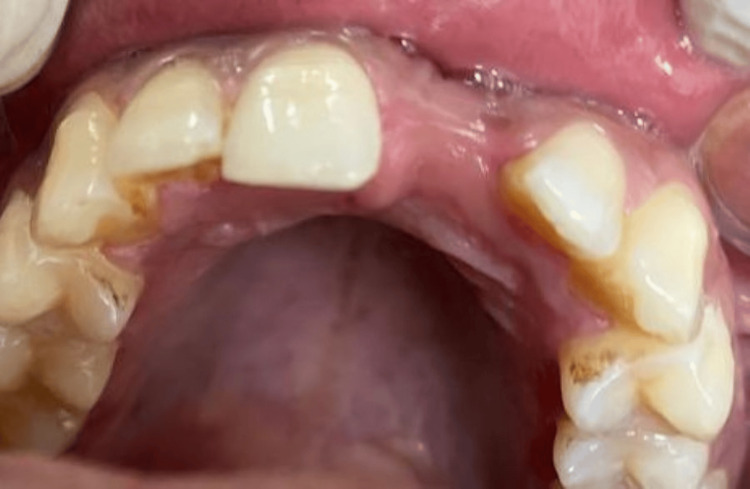
Preoperative intra-oral image depicting narrow alveolar bone with buccal concavity and adequate soft tissue thickness in relation to missing 21.
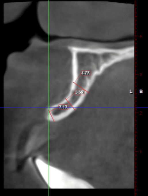
Coronal section of CBCT image showing 3.13 mm of buccolingual bone at the crest, 3.69 mm bone at the middle third, and 4.77 mm bone apically denoting inadequate buccolingual bone for implant placement using conventional osteotomy.
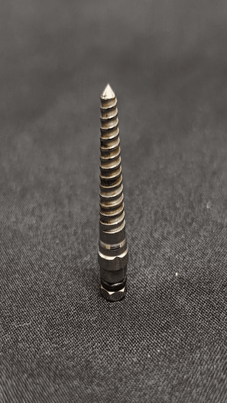
Bone expansion screw/thread former used for lateral bone expansion in narrow ridges or ridges with buccal concavity, which is used sequentially with increasing diameters from 1.6 to 2.8 mm and 1.9 to 3.4 mm.
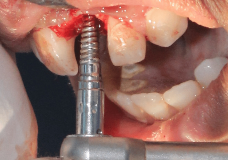
Lateral bone expansion done using bone expansion screws of increasing diameters with the help of a ratchet that allows half of a turn at a time.
After assessment of successful osseointegration, the prosthetic phase was done, and the implant with respect to 21 was loaded using a cement-retained crown as the screw access hole was labial.
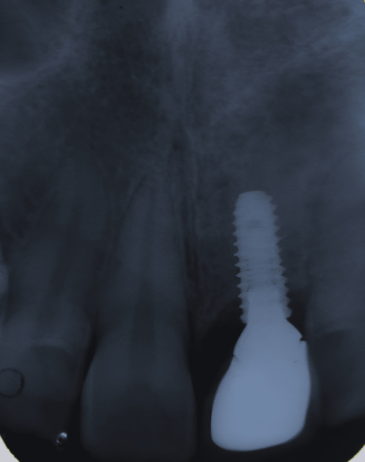
Intra-oral periapical radiograph was taken for the patient at the follow-up visit after one year, showing mild bone loss of 0.2 mm, which is the expected amount of bone loss according to Albrektsson’s criteria.
Socket shield technique
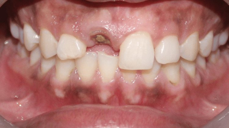
Preoperative intra-oral image showing fractured 11 at the level of cementoenamel junction, with adequate soft tissue thickness but lacking the necessary ferrule for salvaging the tooth using post and core.
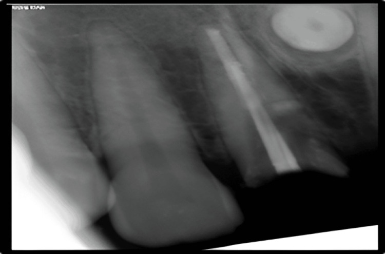
Preoperative RVG image showing root canal treated 21 fractured at CEJ.
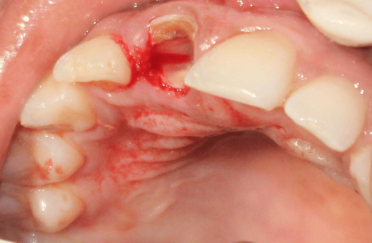
Intra-oral image showing vertical sectioning of the root into buccal and palatal fragments using a long shank bur (Jull Dent-094A) of length 28 mm in a high-speed handpiece.
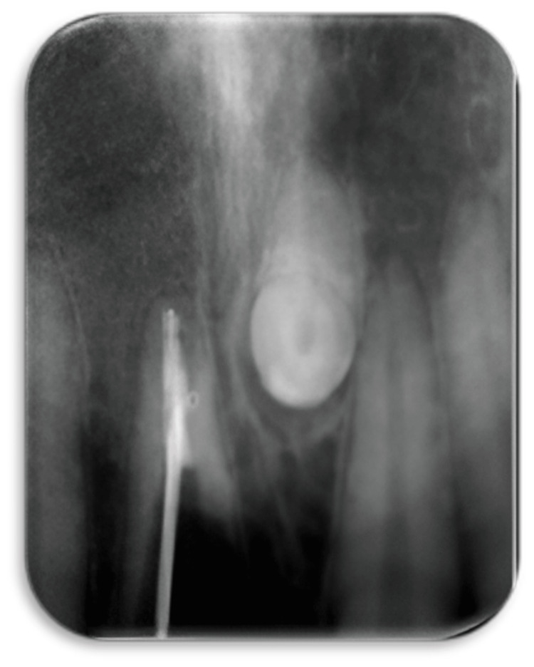
RVG taken intra-operatively to assess the level of root resection.
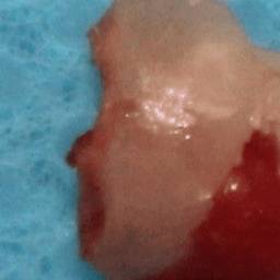
After vertical sectioning, the palatal fragment along with the apex of tooth 11 was extracted with the help of periotome to preserve the buccal root and buccal bone.
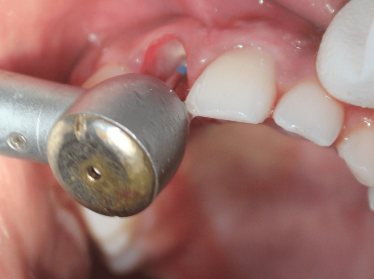
The remaining labial shield was reduced coronally to 1 mm above the alveolar crest using a round bur in a high-speed handpiece, and bevel preparation was done
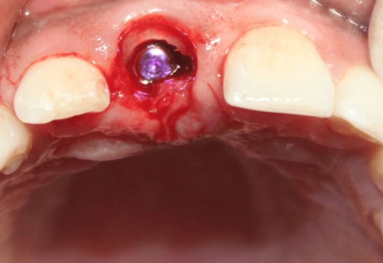
Osteotomy was then sequentially prepared, and a 3.5 x 13 mm implant was inserted palatal to the socket shield.
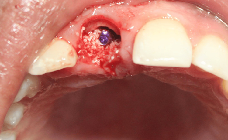
The jump gap between the implant and the shield was filled with 100 mg of osseograft (xenogenic particulate bone graft), which is a DMBM.
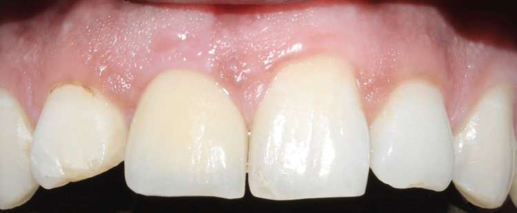
Implant in relation to 11 was loaded after a period of eight months with a cement-retained crown.
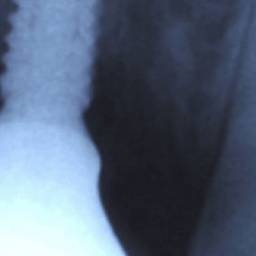
During the follow-up visit one year after loading, IOPA was taken, which shows the intact buccal shield and a negligible amount of bone loss in relation to 11.
Osseodensification
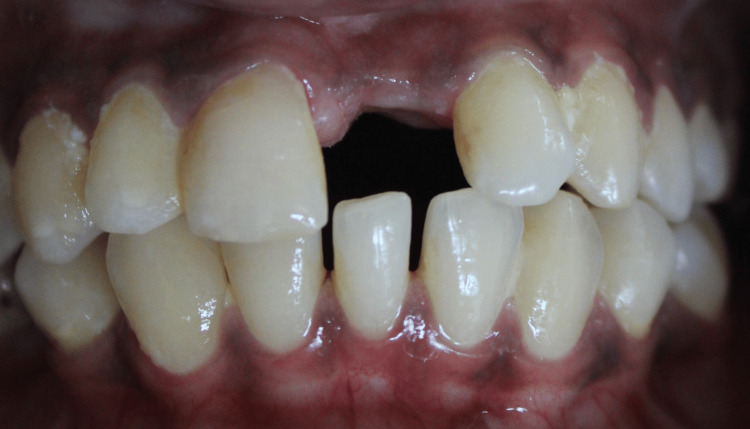
Preoperative intra-oral image showing narrow edentulous space with respect to missing maxillary left central incisor with an adequate soft tissue thickness.
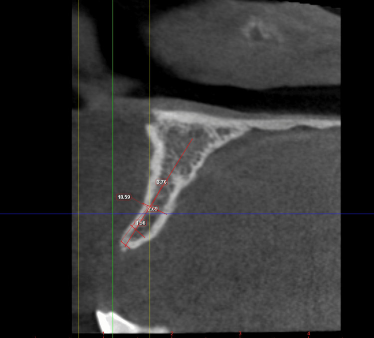
The coronal section of the CBCT image of the patient showing 1.56 mm B-L width at the crest, 2.69 mm in the middle third, and 3.76 mm at the apical region of 21 region, which is inadequate for implant placement using conventional osteotomy.
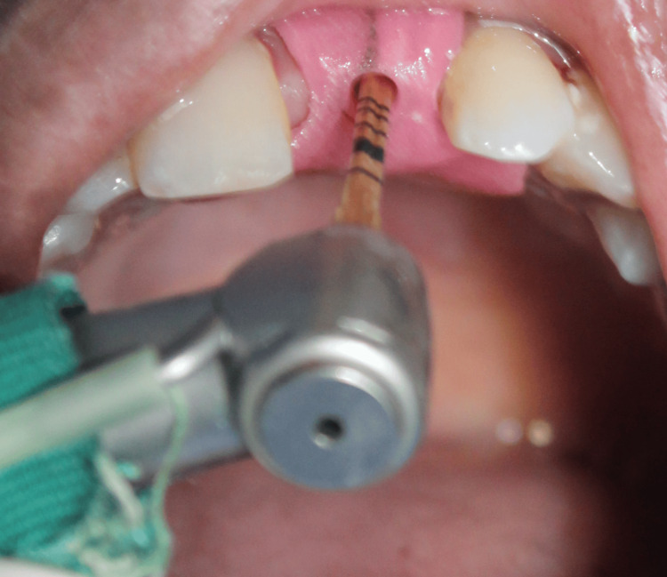
Densah burs are used in counterclockwise rotation for the preparation of osteotomy to achieve bone condensation and improve primary stability.

Image depicting densah bur, which has more land than conventional straight drills that helps in controlled plastic deformation of bone.
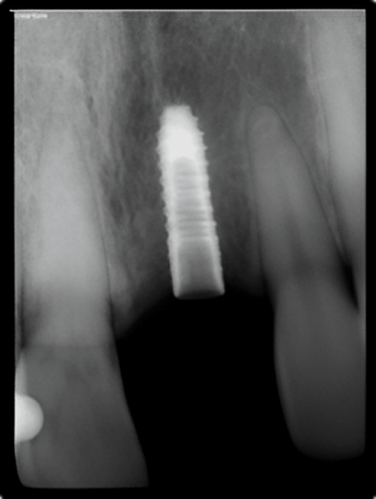
Immediate postoperative IOPA depicting bone condensation with respect to 21.
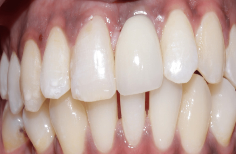
Customized abutment was fabricated and torqued for 20 Ncm followed by luting of a cement-retained crown on the implant with respect to 21.
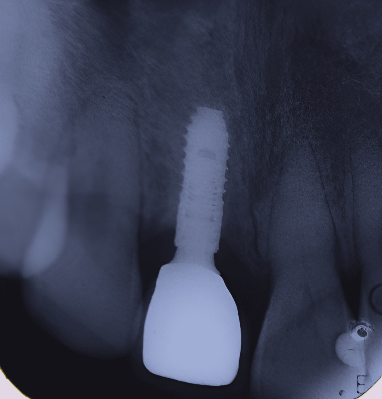
IOPA taken during follow-up visit after six months showing successful functioning of the prosthesis without bone loss.
Resources
-
Real-World Implant Survival in the Esthetic Zone: Uncovering Timing-Specific Risk Patterns in a Large Cohort. Doron Haim Hadar Better Isabelle Meinster et al., Clinical oral implants research. Volume: , Issue: , 2026
-
Long-term outcome of implants exhibiting buccal soft tissue dehiscence in the esthetic zone. Francisco J Enrile de Rojas Javier Ventura de la Torre José Miguel Romero Griñolo et al., Journal of esthetic and restorative dentistry : official publication of the American Academy of Esthetic Dentistry … [et al.]. Volume: 36, Issue: 4, 2024
-
Contemporary Rationales for Cemented Implant Restorations in the Esthetic Zone. Young K Kim Julián Conejo David J Wagner Eduardo Britton Todd R Schoenbaum et al., Journal of esthetic and restorative dentistry : official publication of the American Academy of Esthetic Dentistry … [et al.]. Volume: , Issue: , 2024
-
Guidelines for Immediate Vs Delayed Dental Implant Placement in the Esthetic Zone. Joseph Carpentieri Gary Greenstein et al., Compendium of continuing education in dentistry (Jamesburg, N.J. : 1995). Volume: 45, Issue: 7, 2024 Jul-Aug
-
Enhancing Anterior Esthetic Zone Implant Placement Through Bone Manipulation Techniques: A Case Series. Indu Raj Harinee A Anjana S Raj Arvind K Uikey Femitha Syed et al., Cureus. Volume: 16, Issue: 7, 2024
-
Failure Rate of Dental Implants in the Esthetic Zone: A Systematic Review and Meta-Analysis. Manar Alzahrani Sondus Bakhreibah Nada Alharbi Lama Alamoudi Seba Halloul Sara Alamoudi Raghad Alharthi Salem Baghdadi Ahmed Alamoudi et al., Cureus. Volume: 16, Issue: 7, 2024
-
Dental implant placement with simultaneous localized ridge augmentation using L-shaped titanium mesh in the esthetic zone: a case report. Ziad Albash Eva Hnaino Ali Khalil et al., Journal of surgical case reports. Volume: 2024, Issue: 3, 2024
-
Fixed screw-retained interim restorations with immediate implant placement in esthetic zone: A case series with six different techniques. Udatta Kher Pravinkumar G Patil Ali Tunkiwala Smita Nimbalkar et al., Journal of Indian Prosthodontic Society. Volume: 22, Issue: 1, 2022 Jan-Mar
-
Resorbable Membrane Pouch Technique for Single-Implant Placement in the Esthetic Zone: A Preliminary Technical Case Report. Akiyoshi Funato Chihiro Ishikura Koji Naito Akira Hasuike et al., Bioengineering (Basel, Switzerland). Volume: 9, Issue: 11, 2022
-
Emergence Contours for Single-Unit Implant Provisionals in the Esthetic Zone. Todd R Schoenbaum Young K Kim Faris Khalifa et al., Compendium of continuing education in dentistry (Jamesburg, N.J. : 1995). Volume: 42, Issue: 7, 2021 Jul-Aug
-
Improved volume and contour stability with thin socket-shield preparation in immediate implant placement and provisionalization in the esthetic zone. Serhat Aslan et al., The international journal of esthetic dentistry. Volume: 13, Issue: 2, 2018
-
Vertical ridge augmentation in the esthetic zone. Isabella Rocchietta Luca Ferrantino Massimo Simion et al., Periodontology 2000. Volume: 77, Issue: 1, 2018