New studies on the long-term survival rates of dental implants and failure risk factors, always catch our attention. So This Week in Dental Implants, we wanted to highlight a new paper that evaluated Laser-Lok ® Dental Implants over a 20-year period. 1
About Laser-Lok
Laser-Lok ®, from Biohorizons, is a surface treatment applied to dental implants that involves creating microgrooves or channels (size 8 μm) on the implant surface using laser technology. The Laser-Lok surface is designed to enhance the attachment and growth of soft tissue, such as gingival fibroblasts, around the implant.
Specifically, the laser-textured surface features monodirectional mesoscale channels and woven oblique microridges, which promote uniform cell growth and increased resistance to detachment. This surface treatment has been shown to improve the retention and proliferation of fibroblasts, as well as upregulate adhesion proteins like fibronectin and integrin β-1. These characteristics contribute to better soft tissue integration and stability around the implant, potentially leading to improved clinical outcomes. 2
Study Results with the Seven Risk Factors
Overall the study results were positive and seven risk factors for failures were documented.
Between January of 2000 and December of 2005, 730 LL implants were placed in 312 patients for a variety of clinical indications. Following implant placement, patients were checked at follow-up visits every six months for an average of 16.8 years. The Kaplan-Meier cumulative survival rate (CSR) at 5, 10, 15, and 20 years were 98.2%, 97.1%, 95.8%, and 95.3% respectively. Seven factors were identified associated with late implant failures:
- male,
- smoking,
- maxillary posterior area,
- immediate dental implant placement
- immediate loading protocol,
- (D4) bone quality, and
- ≥ 35 Ncm insertion torque. 1
Case Studies
Both Cases studies below are provided by: Spinelli A, Zamparini F, Romanos G, Gandolfi MG, Prati C. Tissue-Level Laser-Lok Implants Placed with a Flapless Technique: A 4-Year Clinical Study.
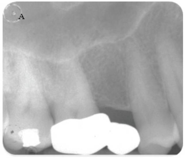
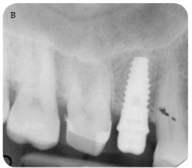
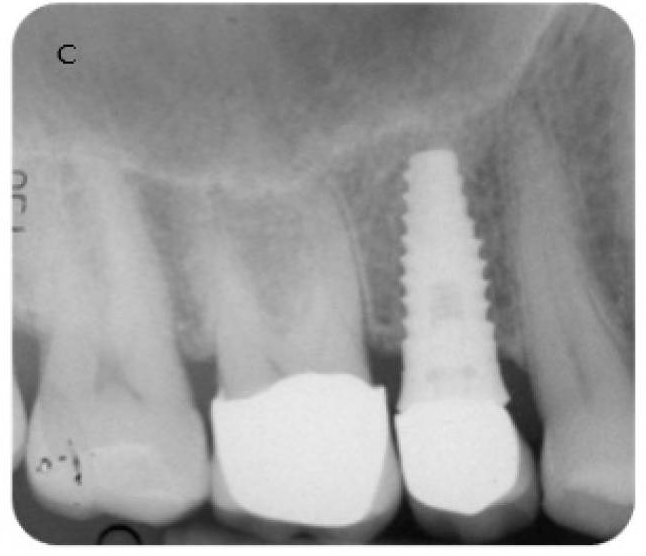
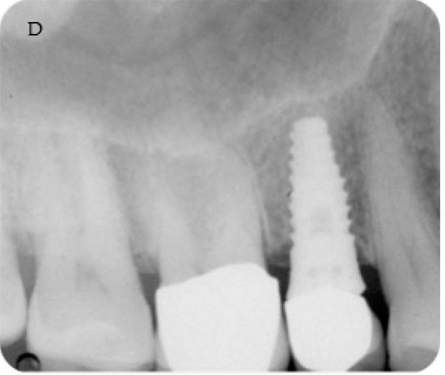
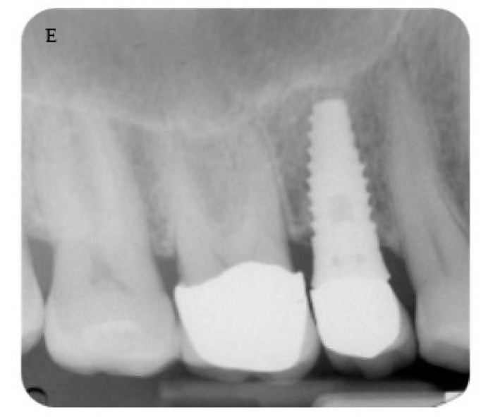
A 4.6 mm tissue-level Laser-'Lok implant was placed with a flapless approach. (A ) Pre-operative radiograph. (B ) Periapical radiograph shows the abutment screwed and the cemented provisional crown. Follow-up at 3 months. (C ) Follow-up at 12 months with the metal ceramic crown cemented on the abutment. MBLs were stable at (D ) 36 months and (E ) 48 months, with limited bone loss.
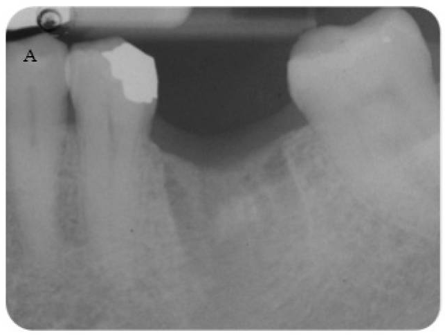
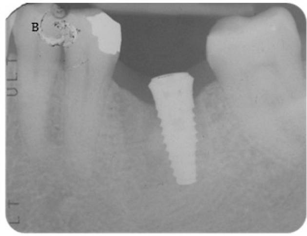
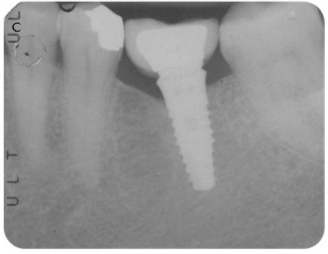
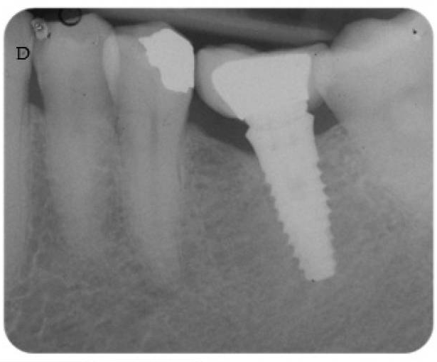
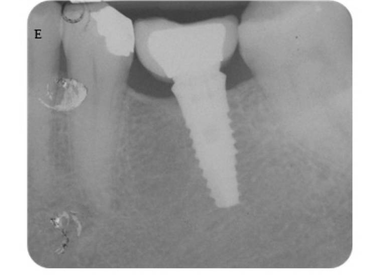
(A ) Early timing placement to rehabilitate a single edentulous ridge. Tooth was extracted 2 months before for a root fracture. (B ) Implant (Laser-Lok) placement with a flapless approach. Please note that the neck was positioned at tissue level. (C ) Periapical radiograph after abutment and definitive crown cementation. (D ) Follow-up at 36 months and (E ) 48 months showed MBL stability around the implant.
The use of Laser-Lok implants placed at tissue level using a flapless technique is supported by this four-year clinical research. During the 48-month follow-up, minimal bone loss and stable marginal bone levels were seen. These values were in line with those reported in the literature. This transmucosal flapless technique allows a soft tissue healing as demonstrated by periodontal parameters that were stable during the 4 years of follow-up. No signs of inflammation or soft tissue alteration around the laser-microtextured neck were observed. The wider diameter implant seems to preserve bone levels. 3
References
-
Guarnieri R, Reda R, Zanza A, Miccoli G, Abbagnale R, Di Nardo DD, Testarelli L. Long-Term (> 20 years) Evaluation and Analysis of Failure Risk Factors of Laser-Lok® Dental Implants in Patients Attending a Private Practice Setting: A Retrospective Audit. Int J Oral Maxillofac Implants. 2025 Sep 12;0(0):1-31.
-
Nevins M, Nevins ML, Camelo M, Boyesen JL, Kim DM. Human histologic evidence of a connective tissue attachment to a dental implant. Int J Periodontics Restorative Dent. 2008
-
Spinelli A, Zamparini F, Romanos G, Gandolfi MG, Prati C. Tissue-Level Laser-Lok Implants Placed with a Flapless Technique: A 4-Year Clinical Study. Materials (Basel). 2023 Feb 2;16(3):1293.