A recent commentary prompted us to revisit research comparing hydrophilic SLActive implant surfaces with conventional SLA surfaces. Hydrophilic surfaces are designed to potentially support early bone healing and osseointegration. But is there really a meaningful clinical advantage? A randomized split-mouth clinical trial comparing modified hydrophilic SLActive and conventional SLA implant surfaces found no statistically significant difference in marginal bone-level changes from loading to 12 months. Both surfaces demonstrated favorable short-term clinical outcomes.
Why Hydrophilicity Matters
SLA (sandblasted, large-grit, acid-etched) is a well-documented moderately rough implant surface. SLActive builds on the same basic surface topography but uses modified processing and saline storage to create a hydrophilic surface that interacts more readily with blood and other fluids.
The goal is to promote early osseointegration and potentially facilitate earlier loading. However, whether these biological advantages translate into meaningfully better clinical outcomes remains an important question.
Earlier Research on Hydrophilic Surfaces
Earlier research established a plausible biological advantage for SLActive, but the clinical implications have been less clear.
- Laboratory: Hydrophilic modified SLA increased osteoblast differentiation markers and suppressed osteoclastic differentiation in vitro [8]. The combination of nanostructure and hydrophilicity also gave the strongest protein adsorption and early blood coagulation [7].
- Early healing in minipigs: In a pilot study, hydrophilic and hydrophobic microrough surfaces showed similar healing patterns, with bone-to-implant contact above 50% by day 10. Hydrophilicity produced only a slight tendency toward more bone apposition at day 15 [6].
- Human histology: In volunteers, SLActive showed more bone-to-implant contact at 2 and 4 weeks (48.3% vs. 32.4% at 4 weeks), but the difference was gone by 6 weeks [9]
- Possible advantage of SLActive for Palatal Implants: A 2025 systematic review of six randomized trials involving 326 implants found broadly comparable implant stability between SLA and SLActive during the first 12 weeks in the maxilla and mandible, with a possible advantage for palatal implants. [3]
- Anticoagulated patients: No difference In a split-mouth RCT of 20 patients on anticoagulant therapy (80 small-diameter titanium-zirconium implants), SLActive and SLA showed no significant differences in implant stability (ISQ) at any time point, and both achieved 100% survival and success at one year. [5]
- Biological differences often diminished over six to eight weeks: A 2011 review of laboratory, animal, and clinical evidence found that early biological differences often diminished over six to eight weeks, while both surfaces demonstrated favorable clinical outcomes. [4]
Taken together, the literature suggests that SLActive can influence early biological responses, but whether those differences provide a consistent, meaningful clinical advantage remains uncertain.
The Latest Study: Hydrophilic and Conventional Surfaces Perform Comparably
In this dual-center, randomized split-mouth trial, 68 patients each received one modified SLActive and one conventional SLA implant (136 implants) in healed posterior sites. Examiners were blinded, and the primary outcome was radiographic marginal bone-level change from loading to 12 months (Vílchez et al. 2026 [1]).
Key findings:
- Primary outcome: From loading to 12 months, marginal bone level changed by −0.04 mm around SLActive and −0.07 mm around SLA (p = 0.658). The negative values reflect slight apparent bone gain, with no significant difference between surfaces.
- Overall bone remodeling: From placement to 12 months, changes were 0.20 mm for SLActive vs. 0.06 mm for SLA. This was statistically significant (p = 0.024), with slightly more change around SLActive. The difference was small and clinically negligible, and both surfaces stayed well below the 1.5 mm threshold for concerning bone loss.
- Other clinical outcomes: Implant stability, probing depth, bleeding on probing and wound healing were comparable. No peri-implantitis was observed, and no implants were lost after loading.
Read the original study: Clinical and Radiographic Performance of Two Distinct Sandblasted, Large-Grit, Acid-Etched Implant Surfaces: A Split-Mouth Randomized Clinical Trial
What Does This Mean for Clinicians?
In their commentary, Afrashtehfar and Jurado [2] conclude that both surfaces performed excellently over one year of loading and that hydrophilicity showed no clinically meaningful superiority. The small placement-to-12-month difference was statistically significant, but it did not translate into a clinical advantage for either surface. Surgical and prosthetic factors, such as implant position, depth and abutment design, likely matter more for bone stability than surface chemistry.
Importantly, the results do not establish that hydrophilic surfaces offer no benefit in other situations. This trial enrolled healed, non-augmented sites with conventional loading at about 10 weeks, so it can’t speak to other scenarios. Whether hydrophilicity helps in poor-quality bone or with earlier loading protocols remains an open question that prior reviews say needs further study.
Clinical Images
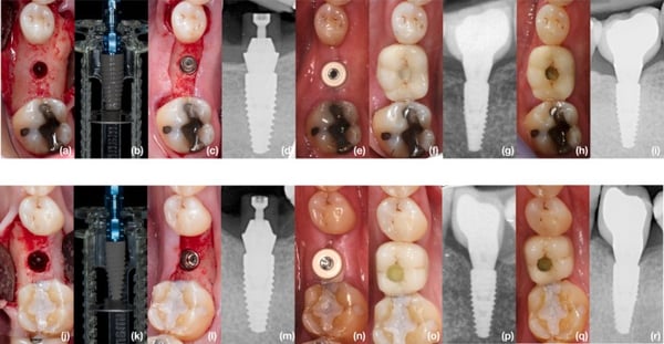
(Source: Vílchez B, Caneiro L et al. 2026 [1])
Clinical and radiographic sequence of implant placement and restoration over 12‐month follow‐up. Images from (a) to (r) correspond to the same patient. (a–i) correspond to the test implant surface (SLActive) and (j–r) correspond to the control implant surface (SLA). (a, j) Implant bed preparations, envelopes opening occurs at this moment. (b, k) Implants prepared to be placed. (c, l) Implants placed approximately 1 mm sub‐crestally. (d, m) Periapical X‐ray after surgery end. (e–n) Digital impression visit. (f–o) Implant loading visit. (g, p) Periapical X‐ray after implant loading. (h–i, q–r) Implants at 12 months follow‐up.
For additional case Photos please see: Clinical and radiographic performance of two distinct sandblasted, large-grit, acid-etched implant surfaces: a split-mouth randomized clinical trial.
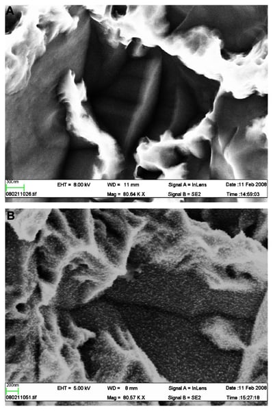
(Source: Wennerberg A, Galli S, Albrektsson T. 2011 [4])
(A) Scanning electron microscopic image of a SLA implant, 80,000× magnification. No distinct nanostructures can be observed. (B) Scanning electron microscopic image of a SLActive implant, 80,000× magnification. Distinct nanostructures can be observed.
For additional case Photos please see: Current knowledge about the hydrophilic and nanostructured SLActive surface.
References
- Vílchez B, Caneiro L, Lima C, Sanz-Sánchez I, Montero E, Figuero E, Blanco J, Sanz M. Clinical and radiographic performance of two distinct sandblasted, large-grit, acid-etched implant surfaces: a split-mouth randomized clinical trial. Clin Oral Implants Res. 2026;37(2):141–154.
- Afrashtehfar KI, Jurado CA. Do hydrophilic implant surfaces provide a clinically meaningful advantage? Evid Based Dent. 2026 Oct 23.
- Patel R, Patel S, Girgis W, Ahmed W, Barrak F. A systematic assessment of the stability of SLA vs. SLActive implant surfaces over 12 weeks. Evid Based Dent. 2025;26(1):67–68.
- Wennerberg A, Galli S, Albrektsson T. Current knowledge about the hydrophilic and nanostructured SLActive surface. Clin Cosmet Investig Dent. 2011 Sep 5;3:59-67.
- Marković A, Đinić A, Calvo Guirado JL, Tahmaseb A, Šćepanović M, Janjić B. Randomized clinical study of the peri-implant healing to hydrophilic and hydrophobic implant surfaces in patients receiving anticoagulants. Clin Oral Implants Res. 2017;28(10):1241–1247.
- Vasak C, Busenlechner D, Schwarze UY, et al. Early bone apposition to hydrophilic and hydrophobic titanium implant surfaces: a histologic and histomorphometric study in minipigs. Clin Oral Implants Res. 2014;25(12):1378–1385.
- Kopf BS, Ruch S, Berner S, Spencer ND, Maniura-Weber K. The role of nanostructures and hydrophilicity in osseointegration: in-vitro protein-adsorption and blood-interaction studies. J Biomed Mater Res A. 2015;103(8):2661–2672.
- Bang SM, Moon HJ, Kwon YD, Yoo JY, Pae A, Kwon IK. Osteoblastic and osteoclastic differentiation on SLA and hydrophilic modified SLA titanium surfaces. Clin Oral Implants Res. 2014;25(7):831–837.
- Lang NP, Salvi GE, Huynh-Ba G, Ivanovski S, Donos N, Bosshardt DD. Early osseointegration to hydrophilic and hydrophobic implant surfaces in humans. Clin Oral Implants Res. 2011 Apr;22(4):349-56.