This Week in Dental Implants we are covering electrolytic cleaning of dental implants (also sometimes referred to as galvanic cleaning). Electrolytic cleaning is a decontamination technique that utilizes an electric field to remove biofilm and contaminants from the surfaces of dental implants. Recently, there was a new study released which examined the effect of this treatment on the mechanical properties of dental implants. The study showed that: These treatments do not affect the corrosion resistance of dental implants, and actually favor their mechanical properties in the long term. 1
Background: What is Electrolytic Cleaning of Dental Implants?

According to the latest research, the primary etiology of peri-implantitis remains a bacterial biofilm. Electrolytic cleaning is a novel approach to decontaminate dental implants without effecting their physical properties. Simply put, the process involves the application of low voltage to the implant and simultaneous application of an electrolyte solution. This helps to disrupt the adhesion forces of the biofilm on the implant surface. Technically speaking, what happens is that electrolysis produces hydrogen cations (H+) which penetrate the biofilm. Due to deoxidation, hydrogen bubbles emerge on the implant surface and lift the biofilm off the implant surface.
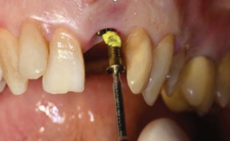
GalvoSurge ® from Straumann is the most widely-known device available on the market that employs the electrolytic cleaning method to clean dental implants. According to the company’s marketing literature:
In just 2 minutes, GalvoSurge® effectively removes biofilm from dental implants and creates optimal conditions for bone regeneration and re-osseointegration.
Note: As of the date of this post, GalvoSurge® is still not available in the US Market. You can contact Straumann directly to inquire about availability.
Case Presentations
Case 1: Treatment of Periimplantitis with Electrolytic Cleaning versus Mechanical and Electrolytic Cleaning: 18-Month Results
Case By: Schlee M, Wang HL, Stumpf T, Brodbeck U, Bosshardt D, Rathe F. Treatment of Periimplantitis with Electrolytic Cleaning versus Mechanical and Electrolytic Cleaning: 18-Month Results from a Randomized Controlled Clinical Trial. J Clin Med. 2021 Aug 6;10(16):3475.


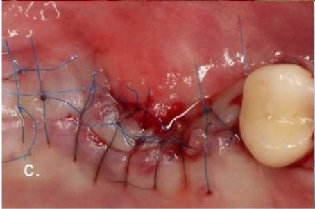
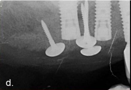

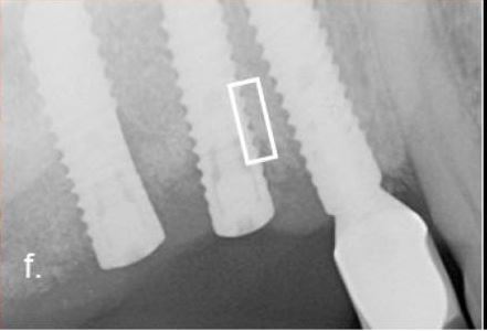

Clinical course of a failed case: (a ) implants 15 and 16 with severe combined intra- and supra-osseous bone defects (RP Class 3); (b ) augmentation after electrolytic cleaning with a combination of mineralized bovine bone mineral and autogenous bone using the Umbrella technique; (c ) tensionless suturing after the crestal mobilization of the flap; (d ) radiograph demonstrating the augmented volume; (e ) clinical situation 6 months after the flap surgery, exposure of the flap caused some loss of augmented volume; (f ) radiograph 11 months after surgery (the implant had to be removed because of suppuration); (g ) histology with a field of interest in the augmented area pointing out regenerated bone and reosseointegration to the implant surface. * Particles of deproteinized bovine bone mineral, +pus.
Case 2: Electrolytic Cleaning and Regenerative Therapy of Peri-implantitis in the Esthetic Area: A Case Report
Case By: Gianfreda F, Punzo A, Pistilli V, Bollero P, Cervino G, D’Amico C, Cairo F, Cicciù M. Electrolytic Cleaning and Regenerative Therapy of Peri-implantitis in the Esthetic Area: A Case Report. Eur J Dent. 2022 Oct;16(4):950-956.

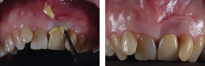
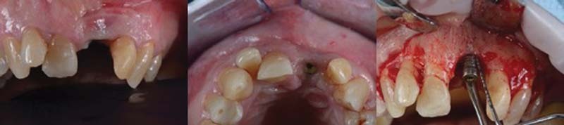
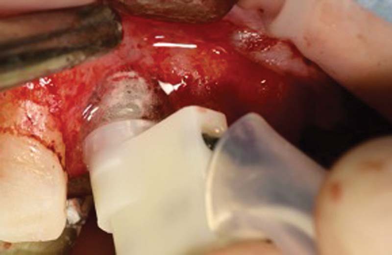


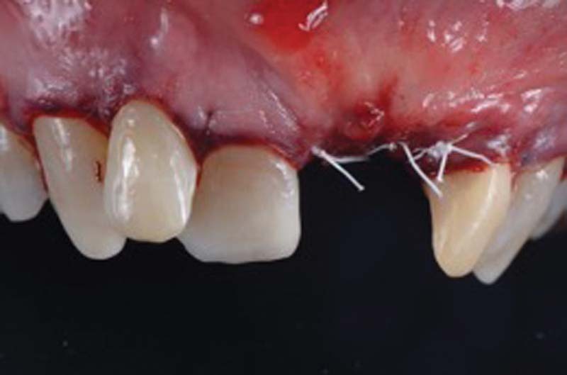
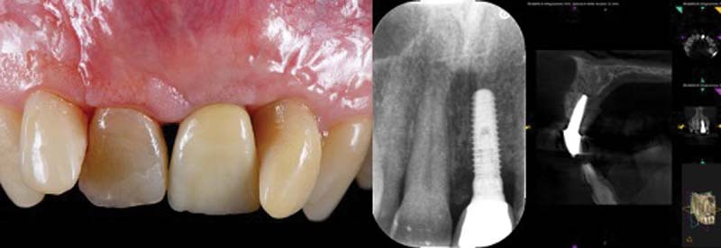
Periapical X-ray showed peri-implant bone resorption and cone-beam computed tomography (CBCT) showed a 5.5-mm circumferential deficit (1). Subsequently, PERIOSTAT gel (iclated doxycycline) is applied, via a carrier with a tip, within the defect (2). After 1 week from decontamination, the crown on implant 2.1 is removed, the cap screw is inserted with application of iodoform paste and then a maryland bridge has been placed (3). Once the soft tissues have healed, after 4 weeks, we can proceed with a regenerative surgery to regenerate the portion of peri-implant bone tissue lost due to peri-implantitis. (4) Once the flap has been removed, the defect degranulated and washed with rifampicin (RIFADIN, Sanofi, Milan, Italy), the implant is decontaminated using an electrolytic approach using GS1000 GalvoSurge Dental AG (5). Once the electrolytic decontamination is completed, the bone defect is filled with a deproteinized bone graft (Bioss, Geistlich Pharma AG, Wolhusen, Switzerland) mixed with autologous bone harvested intraorally and then covered with a resorbable membrane (6). The membrane is then fixed with two metal mini-screws and platelet aggregates are inserted to promote healing (7). Once the regenerative phase is completed, the flap is further passivated through periosteal incisions apical to the flap and then sutured to the anatomic papillae. PTFE 5.0 (Omnia, Fidenza, Italy; mattress suture on the implant area) and PGCL 6.0 (8). The patient is checked every 7 days during the first month and then monthly for 12 months, when a new CBCT is then performed (9). At 12 months, both an endoral periapical control X-ray and a CBCT were performed, showing a complete circumferential filling of the treated peri-implant defect. In fact, a total bone coverage of the shoulder of the 2.1 implant can be seen, with a bone gain of 6 mm on the mesial aspect and 5 mm on the distal aspect.
What Do the Studies Show on Electrolytic Cleaning?
Research indicates that electrolytic cleaning not only achieves significant biofilm removal but also maintains the integrity of the implant surface, avoiding alterations that could compromise its mechanical properties. This makes it a promising option for treating implants affected by peri-implantitis, as it can enhance the potential for successful re-osseointegration and overall implant longevity. Below we have listed a wide range of studies we have found discussing this cleaning technique, including the most recent study we found, mentioned above.
1. Effect Of Electrolytic Cleaning on Mechanical Properties for Titanium Dental Implants with Surface Contamination., Darcio Fonseca , Ramón Pons , Beatriz de Tapia , Alberto Monje , José Nart , Conrado Aparicio , Javier Gil , The International journal of oral & maxillofacial implants. Volume: 0, Issue: 0, 2025
Recently, galvanic cleaning techniques, such as Galvosurge®, which utilize hydrogen formation, have demonstrated significant efficacy in removing biofilm during the decontamination of implant surfaces in the reconstructive therapy of peri-implantitis, This decontamination method avoids the mechanization of the implant and therefore avoids the loss of mechanical properties, loss of good corrosion behavior and avoids the release of particles of different sizes with possible toxic effects, However, the formation of hydrogen on the surface could cause diffusion into the titanium and lead to hydrogen embrittlement of the dental implant, In this study we intend to study the effect of this treatment on the mechanical properties of dental implants, Materials and methods: Ninety dental implants were studied, of which 30 were control, 30 were treated with Galvosurge® and 30 were treated with concentrated hypochlorous acid, The amount of hydrogen in the titanium interior was determined for each of the samples by spectroscopy to elemental analysis of hydrogen TCH600 LECO. Electrochemical corrosion tests were performed on 30 dental implants to determine the corrosion potentials and corrosion intensity for the different treatments. One important factor for the fatigue behavior are the residual stresses which were studied by by Bragg-Bentano X-ray diffraction. Residual stresses were studied by by Bragg-Bentano X-ray diffraction. Fatigue tests were performed using a servohydraulic machine determining the S-N curves by performing triaxial tests (tensioncompression and 5 ̊ torsion) at different loads to simulate human mastication. A study of the fractures of the dental implants was carried out by scanning electron microscopy and the samples were observed by transmission electron microscopy to observe the possible appearance of hydrides in the titanium microstructure, Results: The results showed that the electrolytic technique reduces the presence of hydrogen in the dental implant and the acid treatment increases it causing the presence of hydrides at the grain boundaries of the titanium, It has been shown that galvanic treatments do not affect the corrosion resistance of dental implants. However, attacks with hypochlorous acid increase the corrosion rate due to the acid attack on the titanium surface that favors pitting points. Fatigue tests show that dental implants treated with Galvosurge® have a longer fatigue life than the control due to the lower hydrogen content, It was shown that the increase of hydrogen in the acid-treated implants reduces the fatigue life of the dental implant, Conclusions: This study allows us to conclude that the formation of hydrogen by electrolysis does not cause a diffusion of this element to the titanium nor does it affect corrosion resistance but on the contrary reduces the level of hydrogen which favors its mechanical properties in the long term. Read More2. Electrochemical removal of biofilms from titanium dental implant surfaces., Sebastian Schneider , Michael Rudolph , Vanessa Bause , Andreas Terfort , Bioelectrochemistry (Amsterdam, Netherlands). Volume: 121, Issue: , 2018
The infection of dental implants may cause severe inflammation of tissue and even bone degradation if not treated. For titanium implants, a new, minimally invasive approach is the electrochemical removal of the biofilms including the disinfection of the metal surface. In this project, several parameters, such as electrode potentials and electrolyte compositions, were varied to understand the underlying mechanisms. Optimal electrolytes contained iodide as well as lactic acid. Electrochemical experiments, such as cyclic voltammetry or measurements of open circuit potentials, were performed in different cell set-ups to distinguish between different possible reactions. At the applied potentials of E < -1.4 V, the hydrogen evolution reaction dominated at the implant surface, effectively lifting off the bacterial films. In addition, several disinfecting species are formed at the anode, such as triiodide and hydrogen peroxide. Ex situ tests with model biofilms of E. coli clearly demonstrated the effectiveness of the respective anolytes in killing the bacteria, as determined by the LIVE/DEAD™ assay. Using optimized electrolysis parameters of 30 s at 7.0 V and 300 mA, a 14-day old wildtype biofilm could be completely removed from dental implants in vitro. Read More3. The Effect of In Vitro Electrolytic Cleaning on Biofilm-Contaminated Implant Surfaces., Christoph Ratka , Paul Weigl , Dirk Henrich , Felix Koch , Markus Schlee , Holger Zipprich , Journal of clinical medicine. Volume: 8, Issue: 9, 2019
PURPOSE
Bacterial biofilms are a major problem in the treatment of infected dental and orthopedic implants. The purpose of this study is to investigate the cleaning effect of an electrolytic approach (EC) compared to a powder-spray system (PSS) on titanium surfaces.
MATERIALS AND METHODS
The tested implants (different surfaces and alloys) were collated into six groups and treated ether with EC or PSS. After a mature biofilm was established, the implants were treated, immersed in a nutritional solution, and streaked on Columbia agar. Colony-forming units (CFUs) were counted after breeding and testing (EC), and control (PSS) groups were compared using a paired sample ,-test.
RESULTS
No bacterial growth was observed in the EC groups. After thinning to 1:1,000,000, 258.1 ± 19.9 (group 2), 264.4 ± 36.5 (group 4), and 245.3 ± 40.7 (group 6) CFUs could be counted in the PSS groups. The difference between the electrolytic approach (test groups 1, 3, and 5) and PSS (control groups 2, 4, and 6) was statistically extremely significant (,-value < 2.2 × 10,).
CONCLUSION
Only EC inactivated the bacterial biofilm, and PSS left reproducible bacteria behind. Within the limits of this in vitro test, clinical relevance could be demonstrated. Read More
4. Evidence of re-osseointegration after electrolytic cleaning and regenerative therapy of peri-implantitis in humans: a case report with four implants., Dieter D Bosshardt , Urs R Brodbeck , Florian Rathe , Thomas Stumpf , Jean-Claude Imber , Paul Weigl , Markus Schlee , Clinical oral investigations. Volume: 26, Issue: 4, 2022
OBJECTIVE
To evaluate re-osseointegration after electrolytic cleaning and regenerative therapy of dental implants with peri-implantitis in humans.
MATERIAL AND METHODS
Four dental implants that developed peri-implantitis underwent electrolytic cleaning followed by regenerative therapy with guided bone regeneration. All four implants developed recurrent peri-implantitis and were therefore explanted 6 to 13 months later. Radiographic bone level, probing depth, and bleeding on probing were determined at the time of surgery, 6 months later, and before implant retrieval. The peri-implant tissues were histologically and histomorphometrically analyzed.
RESULTS
All four implants demonstrated radiographic and histological bone gain, reduced probing depth, and bleeding on probing. Radiographic bone gain was 5.8 mm mesially and 4.8 mm distally for implant #1, 3.3 mm and 2.3 mm for implant #2, 3.1 mm and 0.5 mm for implant #3, and 3.5 mm and 2.8 mm for implant #4. The histometric mean and maximum vertical bone gain for implant #1 to #4 was 1.65 mm and 2.54 mm, 3.04 mm and 3.47 mm, 0.43 mm and 1.27 mm, and 4.16 mm and 5.22 mm, respectively. The percentage of re-osseointegration for implant #1 to #4 was 21.0%, 36.9%, 5.7%, and 39.0%, respectively. In one implant, the newly formed bone was deposited directly onto calculus on the implant surface.
CONCLUSIONS
We found that (1) re-osseointegration is possible on a formerly contaminated implant surface and (2) the electrolytic cleaning process seems to be effective enough at sites with calculus residues.
CLINICAL RELEVANCE
Since re-osseointegration can be achieved by electrolytic cleaning, this decontamination technique may be considered as a future treatment concept. Read More
5. Comparison of decontamination efficacy of two electrolyte cleaning methods to diode laser, plasma, and air-abrasive devices., Holger Zipprich , Paul Weigl , Riccardo Di Gianfilippo , Larissa Steigmann , Dirk Henrich , Hom-Lay Wang , Markus Schlee , Christoph Ratka , Clinical oral investigations. Volume: 26, Issue: 6, 2022
OBJECTIVE
To compare the in vitro decontamination efficacy of two electrolytic cleaning methods to diode laser, plasma, and air-abrasive devices.
MATERIAL AND METHODS
Sixty sandblasted large-grit acid-etched (SLA) implants were incubated with 2 ml of human saliva and Tryptic Soy Broth solution under continuous shaking for 14 days. Implants were then randomly assigned to one untreated control group (n = 10) and 5 different decontamination modalities: air-abrasive powder (n = 10), diode laser (n = 10), plasma cleaning (n = 10), and two electrolytic test protocols using either potassium iodide (KI) (n = 10) or sodium formate (CHNaO,) (n = 10) solution. Implants were stained for dead and alive bacteria in two standardized measurement areas, observed at fluorescent microscope, and analyzed for color intensity.
RESULTS
All disinfecting treatment modalities significantly reduced the stained area compared to the untreated control group for both measurement areas (p < 0.001). Among test interventions, electrolytic KI and CHNaO, treatments were equally effective, and each one significantly reduced the stained area compared to any other treatment modality (p < 0.001). Efficacy of electrolytic protocols was not affected by the angulation of examined surfaces [surface angulation 0° vs. 60° (staining %): electrolytic cleaning-KI 0.03 ± 0.04 vs. 0.09 ± 0.10; electrolytic cleaning-CHNaO2 0.01 ± 0.01 vs. 0.06 ± 0.08; (p > 0.05)], while air abrasion [surface angulation 0° vs. 60° (staining %): 2.66 ± 0.83 vs. 42.12 ± 3.46 (p < 0.001)] and plasma cleaning [surface angulation 0° vs. 60° (staining %): 33.25 ± 3.01 vs. 39.16 ± 3.15 (p < 0.001)] were.
CONCLUSIONS
Within the limitations of the present in vitro study, electrolytic decontamination with KI and CHNaO, was significantly more effective in reducing bacterial stained surface of rough titanium implants than air-abrasive powder, diode laser, and plasma cleaning, regardless of the accessibility of the contaminated implant location.
CLINICAL RELEVANCE
Complete bacterial elimination (residual bacteria < 1%) was achieved only for the electrolytic cleaning approaches, irrespectively of the favorable or unfavorable access to implant surface. Read More
6. Use of electrical field for biofilm implant removal., S Bernardi , E Qorri , G Botticelli , A Scarano , G Marzo , R Gatto , A Greco Lucchina , C Mortellaro , E Lupi , C Rastelli , G Falisi , European review for medical and pharmacological sciences. Volume: 27, Issue: 3 Suppl, 2023
OBJECTIVE
New methods for biofilm removal are being investigated. A recent new one involves the use of the electric field for biofilm removal. In particular, electrolytic cleaning works on the adhesion forces of the biofilm on the surfaces, with few studies showing promising results in decontamination and implant re-integration in the bone. This study aims at assessing the effect of a new decontamination device that implies the electric field for implant-biofilm removal.
MATERIALS AND METHODS
Three implants affected by peri-implantitis were selected for the study. After the treatment, the implants were observed by the Scanning Electron Microscopy.
RESULTS
All three samples showed no microbial biofilm in the application area, while the rest of the surface observed was covered with microbial biofilm, with an intensely thickened bacterial population.
CONCLUSIONS
Peri-mucositis and peri-implantitis prevention and early treatments are essential for implant maintenance, thus saving the surrounding hard and soft tissues. The technological innovation is providing electrolytic devices which act not only on the microbial population but on the biofilm adhesion to the implant surface, with promising results for a new and valid therapeutic option. Read More
7. Dental Implant Surface Decontamination and Surface Change of an Electrolytic Method versus Mechanical Approaches: A Pilot In Vitro Study., Mariana Anselmo Assunção , João Botelho , Vanessa Machado , Luís Proença , António P A Matos , José João Mendes , Lucinda J Bessa , Nuno Taveira , Alexandre Santos , Journal of clinical medicine. Volume: 12, Issue: 4, 2023
Dental implants are the preferred fixed oral rehabilitation for replacing lost teeth. When peri-implant tissues become inflamed, the removal of plaque accumulating around the implant becomes imperative. Recently, several new strategies have been developed for this purpose, with electrolytic decontamination showing increased potential compared to traditional mechanical strategies. In this in vitro pilot study, we compare the efficacy of an electrolytic decontaminant (Galvosurge) with an erythritol jet system (PerioFlow) and two titanium brushes (R-Brush™ and i-Brush™) in removing PAO1 biofilms from implants. Changes in the implant surface after each approach were also evaluated. Twenty titanium SLA implants were inoculated with and then randomly assigned to each treatment group. After treatment, decontamination efficacy was assessed by quantifying colony-forming units (log10 CFU/cm) from each implant surface. Scanning electron microscopy was used to analyse changes in the implant surface. With the exception of R-Brush, all treatment strategies were similarly effective in removing from implants. Major surface changes were observed only in implants treated with titanium brushes. In conclusion, this pilot study suggests that electrolytic decontamination, erythritol-chlorhexidine particle jet system and i-Brush™ brushing have similar performance in removing biofilm from dental implants. Further studies are needed to evaluate the removal of more complex biofilms. Titanium brushes caused significant changes to the implant surface, the effects of which need to be evaluated. Read More8. Effect of chemical and electrochemical decontamination protocols on single and multiple-used healing abutments: A comparative analysis of contact surface area, micro-gap, micro-leakage, and surface topography., Thiha Tin Kyaw , Ahmed Abdou , Sirida Arunjaroensuk , Hidemi Nakata , Manabu Kanazawa , Atiphan Pimkhaokham , Clinical implant dentistry and related research. Volume: 25, Issue: 6, 2023
INTRODUCTION
Although the combined use of chemical and electrochemical decontamination protocols can completely remove contaminants from the surfaces of one-time used healing abutments (HAs), their effectiveness in multiple-used HAs remains unknown. We aimed to investigate the effect of reused HAs frequency on the implant-HA contact surface area, micro-gap, microleakage, and surface topography following chemical and combined chemical and electrochemical decontamination protocols.
METHODS
Ninety bone level titanium implants were assembled with 90 bone level HAs, in which 80 contaminated HA samples were collected from human participants. The retrieved HAs were randomly divided into two groups according to the cleaning protocol: ultrasonication with 5.25% NaOCl solution for 15 min and steam autoclaving (group I); ultrasonication with 5.25% NaOCl solution for 15 min, followed by electrochemical cleaning and steam autoclaving (group II). The control group (group III) comprised 10 new unused HAs. The cleaning protocol was applied after each insertion as follows: (a) single-use and cleaning, (b) double-use and double cleaning cycles, (c) triple-use and triple cleaning cycles, and (d) more than triple-use and more than triple cleaning cycles. The contact surface area and micro-gap were assessed with micro-computed tomography scanning technique, microleakage test using 2% methylene blue staining, surface morphology with scanning electron microscopy, and surface elemental composition with energy-dispersive X-ray spectroscopy analysis.
RESULTS
Group Id exhibited the smallest contact surface area. The values of the micro-gap volumes and microleakage were significantly different (p < 0.001) in the descending order of Id > Ic > Ib > IId > Ia, IIa, and III. Morphological evaluation of Groups IIa, IIb, and IIc revealed that residual biological debris was optimally removed without altering their surface properties.
CONCLUSIONS
Chemical and electrochemical decontamination protocols are more effective than NaOCl cleaning methods, particularly for multiple consecutive uses with better decontamination levels, which decreases micro-gap volume and microleakage without surface alterations. Although the use of combined decontamination protocols for the contact surface area at the implant-HA interface showed comparable results with the control, change in the contact surface area was observed following the NaOCl cleaning methods. Therefore, titanium HA reuse can be considered in multiple times, if they are cleaned and sterilized using combined chemical and electrochemical decontamination protocols. Read More
9. The biofilm removal effect and osteogenic potential on the titanium surface by electrolytic cleaning: An in vitro comparison of electrolytic parameters and five techniques., Yun Zhu , Yangbo Xu , Zhaoting Ling , Congrui Zhao , Antian Xu , Fuming He , Clinical oral implants research. Volume: 35, Issue: 4, 2024
OBJECTIVES
To determine the optimal current and time of electrolytic cleaning (EC), compare its biofilm removal effect with generic treatments and evaluate the influence of EC to surface characteristics and osteogenic potential of SLA titanium (Ti) discs.
MATERIALS AND METHODS
The six-species biofilm-covered Ti discs were placed as cathodes in physiologic saline and subjected to various current and time treatments. The residual biofilms were evaluated to determine the optimal parameters. The contaminated Ti discs were randomized and treated by rotating Ti brush; ultrasonic-scaling with metal tips; ultrasonic-scaling with PEEK tips; air-polishing and EC. The residual biofilms were compared using a lipopolysaccharide kit (LPS), scanning electron microscope (SEM), confocal laser scanning microscopy and colony-forming unit counting. Non-contaminated Ti discs were treated and characterized. The bone marrow mesenchymal stem cells (BMSCs) were cultured on treated non-contaminated Ti discs. The adhesion, proliferation, alkaline phosphatase (ALP) activity and osteocalcin level of BMSCs were assessed.
RESULTS
The parameters at 0.6A5min were considered optimal. For LPS and SEM, EC promoted a significantly greater biofilm removal than the other groups. There were no changes in the Ti discs' colour, topography, roughness and chemical elements after EC, and the electrolysis-treated Ti discs obtained a super-hydrophilic surface. EC positively impacted the proliferation and ALP activity of BMSCs, surpassing the efficacy of alternative treatments.
CONCLUSIONS
EC achieves a near-complete eradication of contaminants on the SLA surface, causes no surface damage with improved hydrophilicity, and promotes the early osteogenic response of BMSCs, which makes it a promising treatment for peri-implantitis. Read More
10. Efficacy of Decontamination Methods for Biofilm Removal from Dental Implant Surfaces and Reosseointegration: An AAP/AO Systematic Review on Peri-implant Diseases and Conditions., Andrea Ravidà , Debora R Dias , Robert Lemke , Paul S Rosen , Martinna M Bertolini , The International journal of oral & maxillofacial implants. Volume: , Issue: 4, 2025
PURPOSE
To evaluate the nonclinical evidence concerning the efficacy of different decontamination methods in facilitating reosseointegration, eliminating biofilm from implant surfaces, and their potential to induce adverse surface modifications and release of material remnants.
MATERIALS AND METHODS
Systematic electronic and manual searches were conducted to identify publications involving animal or human block biopsies, ex vivo/in situ studies, and in vitro studies. Mechanical, chemical, and electrolytic methods for implant decontamination were presented in a descriptive analysis.
RESULTS
A total of 121 studies were included, namely 46 involving animal/human biopsies, 39 ex vivo/in situ experiments, and 36 in vitro investigations. No modality demonstrated significant superiority in terms of reosseointegration outcomes. Ex vivo, in situ, and in vitro studies reported that greater biofilm removal from implant surfaces occurred with polyetheretherketone (PEEK) ultrasonic tips, air-powder abrasive (APA), erbium: yttrium-aluminum-garnet (Er:YAG) laser, and electrolytic cleaning. Minimal surface alterations were noted with soaked cotton pellets, APA, specific settings of Er:YAG laser, erbium, chromium: yttrium-scandium-gallium-garnet (Er,Cr:YSGG) laser, electrolytic treatment, and cold atmospheric plasma. Titanium or stainless steel curettes, ultrasonic tips, titanium brushes, and implantoplasty induced significant surface alterations and peak flattening of implant threads. Plastic and carbon curettes as well as PEEK ultrasonic tips and APA left material remnants.
CONCLUSIONS
Implant reosseointegration is possible following appropriate surface decontamination. Application of Er:YAG laser, electrolytic cleaning, and APA stand out as the methods that most closely embody the ideal characteristics of an effective decontamination protocol. Read More