This Week in Dental Implants we wanted to focus on two new interesting studies pertaining to the survival of dental implants. The first study focused on survival rates of implants placed in previously failed sites, while the second addressed the different outcomes for tissue‐level (TL) and bone‐level X (BLX) implants. With regards to reimplantation at a failed site, we wanted to share an earlier case study that focused on the potential of bruxism and sinus infection as factors for repeated failures.
Survival and Risk Factors of Implants Placed in Sites of Previous Failures
This large retrospective study 1 analyzed 177,410 dental implants placed in 59,238 patients between 2014 and 2023 to evaluate the success of implants placed in sites where previous implants had failed.
Overall implant survival remained excellent at 97.5% after an average follow-up of 4.6 years. However, success rates declined with each additional implant placed at the same site. First implants had a survival rate of 97.6%, while second implants dropped to 92.2%, and third or subsequent implants declined further to 88.5%.
Compared with first-time implants, second implants were 4.6 times more likely to fail, while third or later implants had a 5.7-fold higher risk of failure. The increased risk was especially pronounced during the early healing phase, where second implants were nearly 7 times more likely to fail and third or later implants were more than 9 times as likely to fail.
The study also identified several independent risk factors for implant failure, including:
- Smoking
- Male sex
- Penicillin allergy
- Narrow-diameter implants
- Maxillary implant placement
- Short implants
Comparing Tissue‐Level (TL) and Bone‐Level X (BLX) Survival, and Complication Rates
This retrospective study 2 compared the clinical and radiographic performance of 73 tissue-level (TL) and 73 bone-level X (BLX) implants over a 3-year period.
Both implant systems demonstrated excellent outcomes. Tissue-level implants achieved a 100% survival rate, while bone-level X implants had a 98% survival rate after three years. Although both performed well overall, tissue-level implants exhibited slightly more favorable peri-implant bone stability. They showed a small gain in mesial bone between years two and three, while BLX implants experienced greater mesial and distal bone loss over the study period.
Technical complications, such as prosthetic chipping or other restorative issues, were also more common with BLX implants. An important finding was that BLX implants surrounded by more than 2 mm of keratinized mucosa experienced less peri-implant bone remodeling, suggesting that an adequate band of keratinized tissue may be particularly beneficial for this implant design.
Case Images for Tissue‐Level (TL) and Bone‐Level X (BLX)
Case Images below are from: Scognamiglio C, Çakmak G, Schimmel M, Perucchi A, Abou-Ayash S. Influence of Implant Type on Clinical and Radiologic Outcomes: A Cross-Sectional Private Practice-Based Analysis.
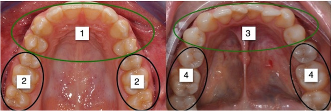
Different implant placement areas used for categorization of the implants. The arch was divided into four regions: anterior superior (teeth 14–24), posterior superior (teeth 15–18 and 25–28), anterior inferior (teeth 34–44), and posterior inferior (teeth 35–38 and 45–48).
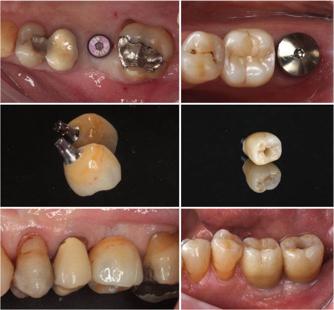
Representative clinical cases of a bone‐level X (BLX) implant and a tissue‐level (TL) implant, respectively.
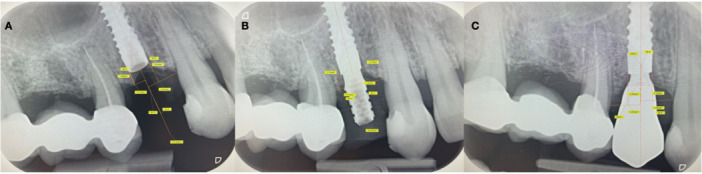
Radiographic bone level measurement for BLX implants. A line was drawn along the central axis of the implant, and a perpendicular reference line was drawn touching the coronal edge of the implant at the mesial and distal points; the vertical distances from this reference line to the bone and to the mesial and distal bone peaks were then measured. (A) Measurement of the implant without a cap screw. (B) Measurement of the implant with a temporary abutment. (C) Measurement of the implant with the final crown.
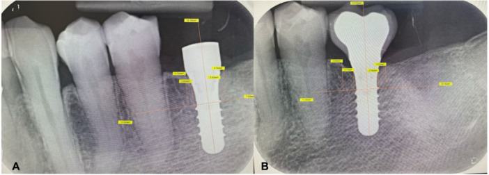
Radiographic bone level measurement for TL implants. A line was drawn along the central axis of the implant, and a perpendicular reference line was drawn touching the most coronal implant thread; the vertical distances from this reference line to the bone and to the mesial and distal bone peaks were then measured. (A) Measurement of the implant with the healing cap. (B) Measurement of the implant with the final crown.
Reasons for Failure with Same Site Implantation: 2 Case Reports
Successful Reimplantation Following Two Implant Failures in a Bruxism Patient
A 68-year-old man underwent immediate implant placement at #46 following extraction of a tooth with a periapical abscess. Although the implant initially integrated successfully, it failed shortly after restoration due to suspected bruxism and excessive occlusal forces. After removal and use of a night guard, a second implant functioned well for over six years before failing for the same reason. A third, wider-diameter implant was then placed and remained stable with good function at the final follow-up. 3
Clinical Takeaway: This case demonstrates that implant reimplantation can be successful after multiple failures when contributing factors—particularly bruxism and occlusal overload—are identified and managed.
Case photos below from Kang DW, Kim SH, Choi YH, Kim YK. Repeated failure of implants at the same site: a retrospective clinical study.
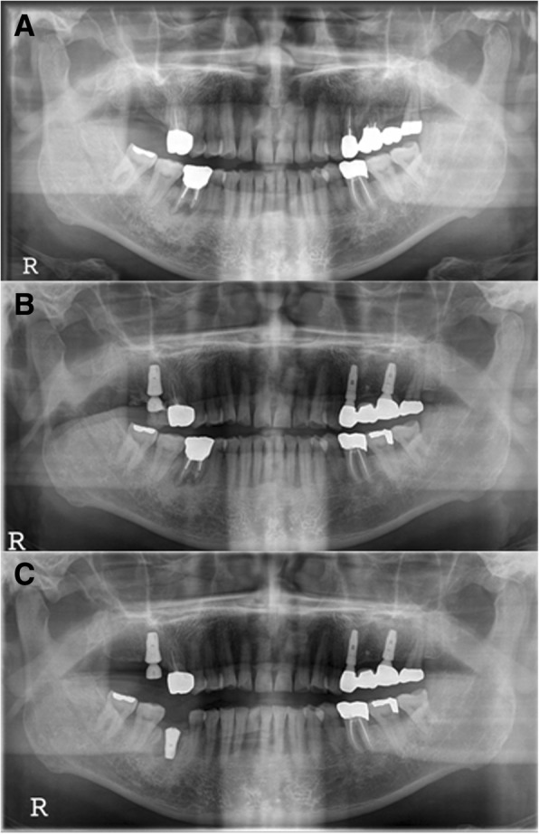
M3, panoramic radiograph, #46i surgery a initial panoramic radiograph, b pre-operative panoramic radiograph, c postoperative panoramic radiograph
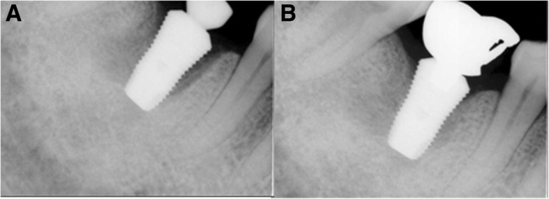
M3, periapical radiograph, a #46i second operation, b #46i prosthesis delivery
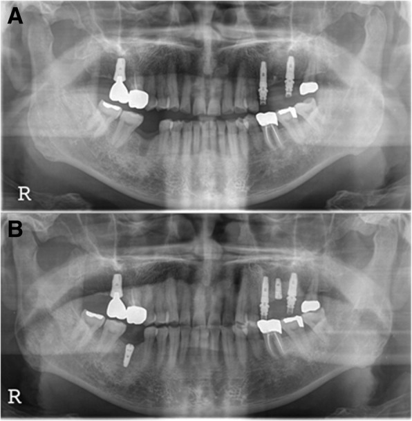
M3, panoramic radiograph, a postoperative 1-month removal of fixture state, b #46i, second installation
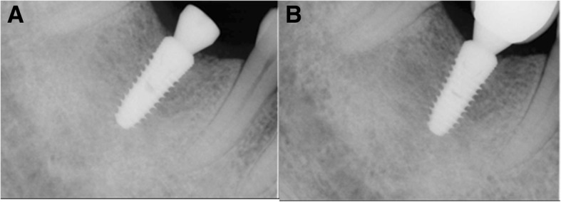
M3, periapical radiograph, a #46i, second operation, b #46i, prosthesis delivery
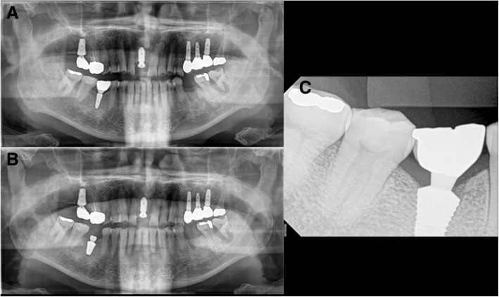
M3, radiographs a #46i second failure, preoperative panoramic radiograph, b #46i, third installation, c #46i, prosthesis delivery
Successful Third Implant After Two Failures in a Grafted Maxillary Sinus
A 62-year-old woman with a history of maxillary sinusitis and severe posterior maxillary bone loss underwent sinus augmentation and implant placement at #27. The first implant failed to osseointegrate and was removed after five months. An immediate replacement implant also failed despite additional grafting. After an 8-month healing period, a third implant was placed using a flapless approach and successfully restored. Although peri-implantitis later developed around the #26 and #27 implants, it was managed with nonsurgical therapy, and both implants remained functional at 4 years and 7 months of follow-up.
Potential Reasons for Failure:
- Previous maxillary sinus disease with possible residual infection.
- Early cover screw exposure, potentially leading to bone graft contamination or loss.
- Implant placement into immature grafted bone, resulting in inadequate osseointegration.
Clinical Takeaway: Even after two failed implants in a compromised grafted maxillary site, successful reimplantation was possible once adequate healing occurred and the site was carefully managed.
Case photos below from Kang DW, Kim SH, Choi YH, Kim YK. Repeated failure of implants at the same site: a retrospective clinical study.
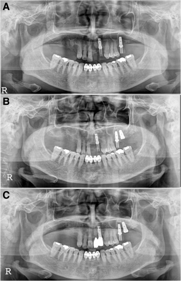
F2, Panoramic radiograph, #27i surgery a initial panoramic radiograph, b postoperative panoramic radiograph, c #27i second operation
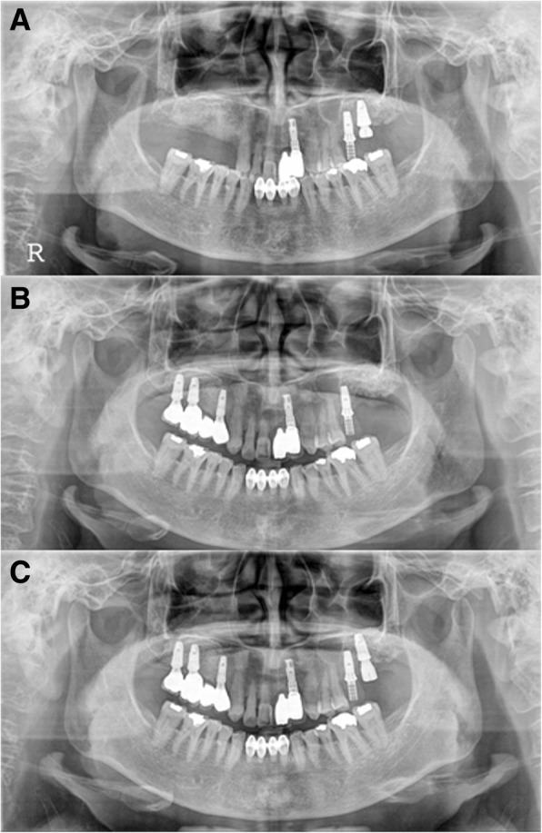
F2, panoramic radiograph, a #27i second installation, b #27i second failure, preoperative panoramic radiograph, c #27i, third installation
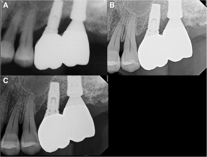
F2, periapical radiograph, a #27i, prosthesis delivery, b #27i peri-implantitis, #26i increasing radiolucent lesion, c #27i peri-implantitis, #26i mesial bone loss
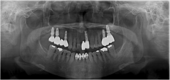
F2, the final panoramic radiograph showing bone loss at #26i and 27i
References
- Asbi T, Haim D, Meinster I, Cahana S, Grecco CM, Better H, Almog R, Zigdon-Giladi H, Shibli JA. Survival and Risk Factors of Implants Placed in Sites of Previous Failures-A Nationwide Retrospective Cohort Study. J Clin Periodontol. 2026 Jul 14.
- Scognamiglio C, Çakmak G, Schimmel M, Perucchi A, Abou-Ayash S. Influence of Implant Type on Clinical and Radiologic Outcomes: A Cross-Sectional Private Practice-Based Analysis. Clin Exp Dent Res. 2026 Aug;12(4).
- Kang DW, Kim SH, Choi YH, Kim YK. Repeated failure of implants at the same site: a retrospective clinical study. Maxillofac Plast Reconstr Surg. 2019 Jul 10;41(1):27.