This Week in Dental Implants we are presenting a case demonstrating a new, and simpler technique for a sinus lift that utilizes ridge spreaders.
Ridge spreaders are specialized surgical instruments that are designed to help in augment narrow edentulous ridges, facilitating the placement of implants by expanding the bone width without the need for additional grafting procedures. This technique is particularly beneficial in cases of horizontal ridge deficiency, as it allows for the preservation of the existing bone and minimizes the need for more complex surgical interventions. In the case below, the authors used a ridge spreader to perform a sinus lift, a very unique use case of ridge spreaders.
As explained in the research article:
In this technical note, we present for the first time, the use of ridge spreaders to safely perform Transcrestal sinus floor elevation (tSFEs). Our simple technique uses existing tools from readily accessible surgical kits from commonly used implant systems. Therefore, this technique can be easily employed in clinical practice. Our technique is minimally invasive and able to successfully perform tSFEs in spite of challenging anatomy. Furthermore, this technique has the advantage of being more comfortable to the patient with no risk of BPPV and no need for instruments to enter the sinus cavity. Read the full article here: Simple technique using ridge spreaders to safely perform a transcrestal sinus floor elevation: a technical note.
> Case photos below provided by: Hue J, Saigo L, Sng TJH, Goh BT. Simple technique using ridge spreaders to safely perform a transcrestal sinus floor elevation: a technical note. J Korean Assoc Oral Maxillofac Surg. 2025 Aug 31;51(4):261-266. doi: 10.5125/jkaoms.2025.51.4.261.
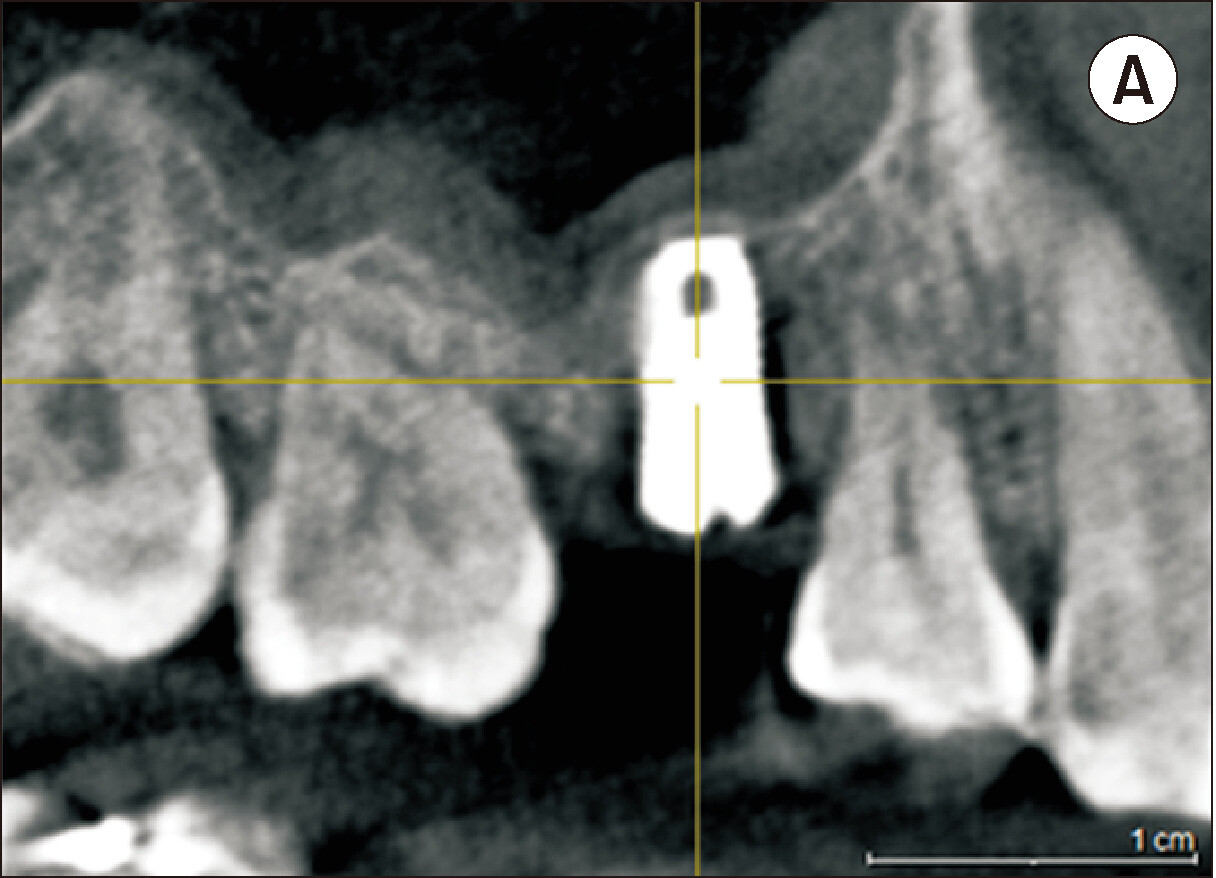
Figure 1
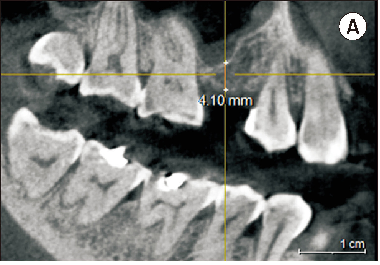
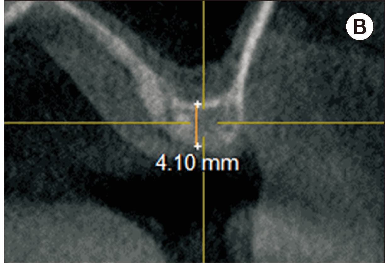
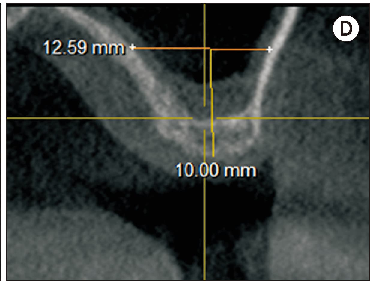
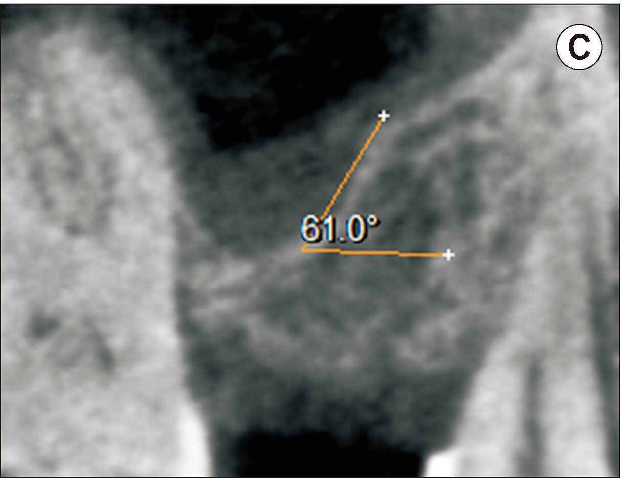
A, B. Pre-operative cone-beam computed tomography (CBCT) showing residual subantral bone height of 4.10 mm. C. Oblique sinus floor morphology at site of planned implant placement. D. CBCT showing buccopalatal sinus width measured at 10.00 mm from the alveolar crest.
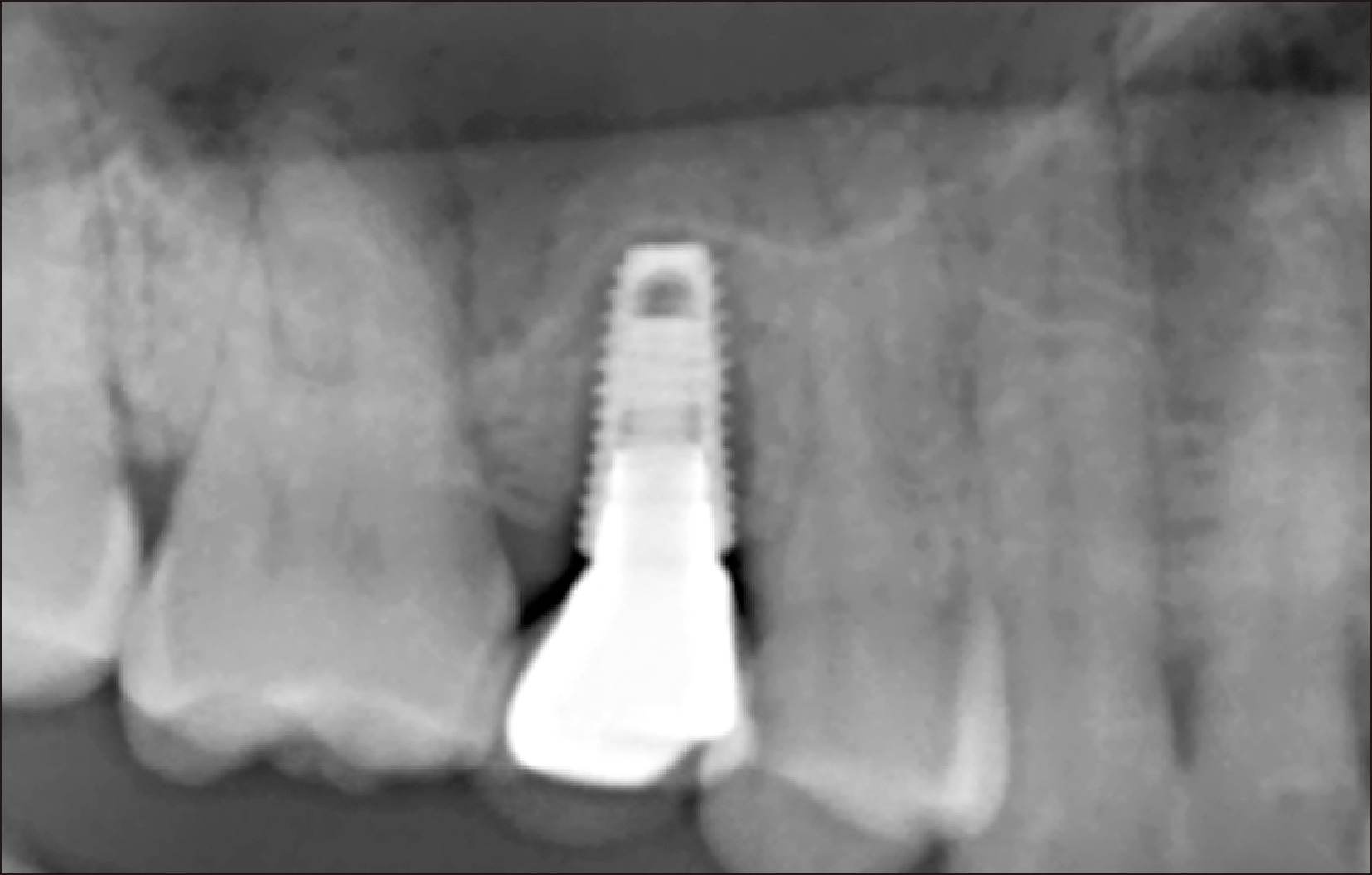
Figure 2
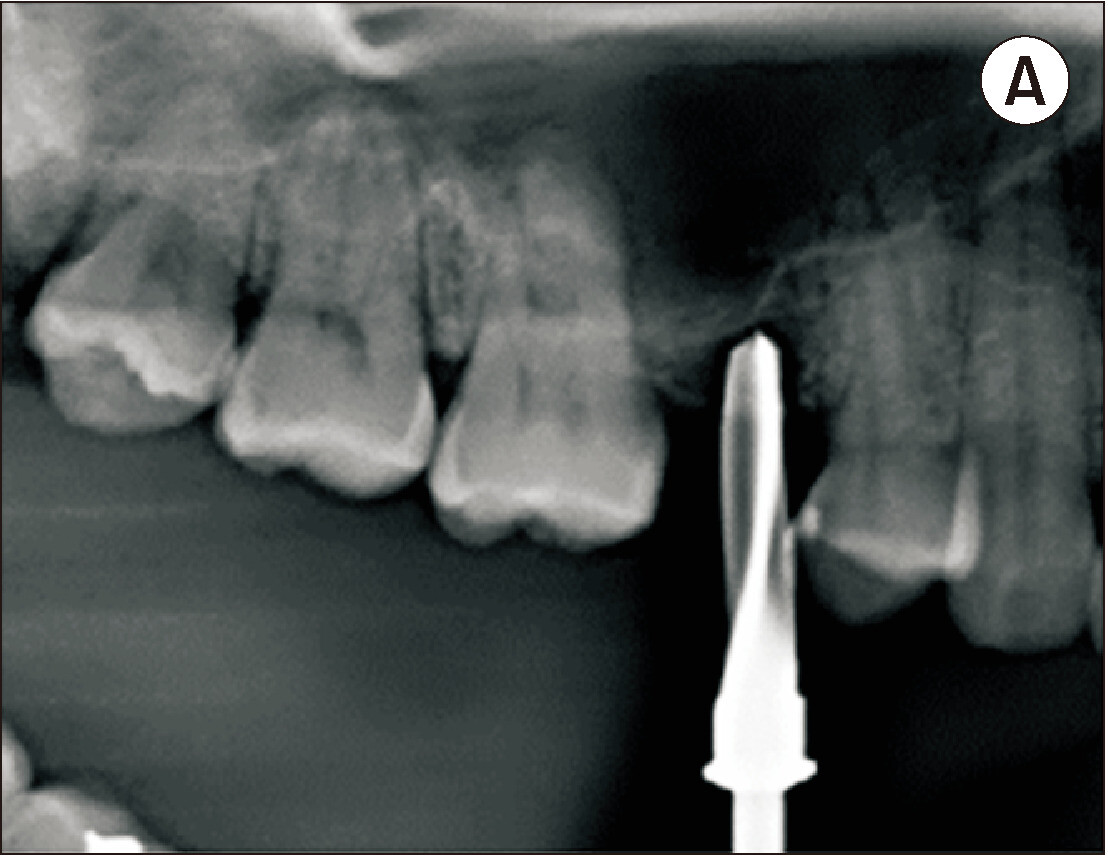
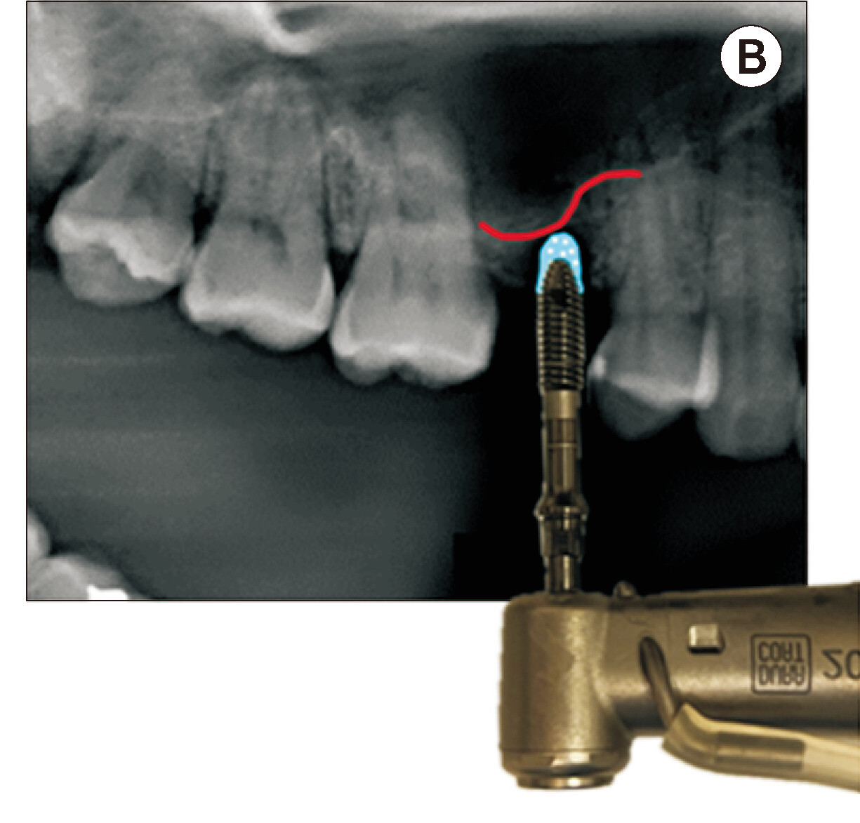
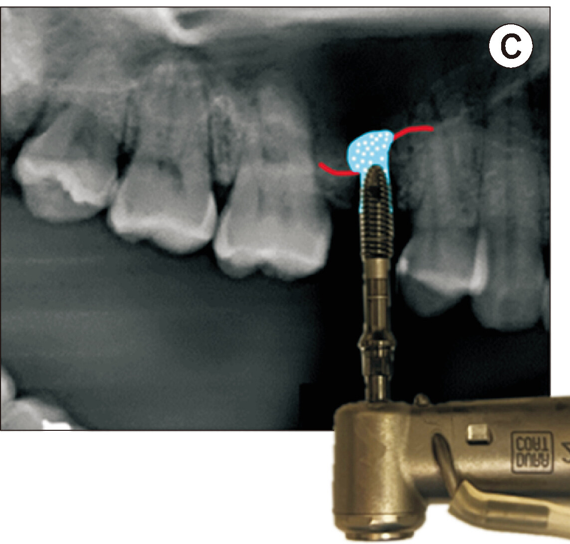
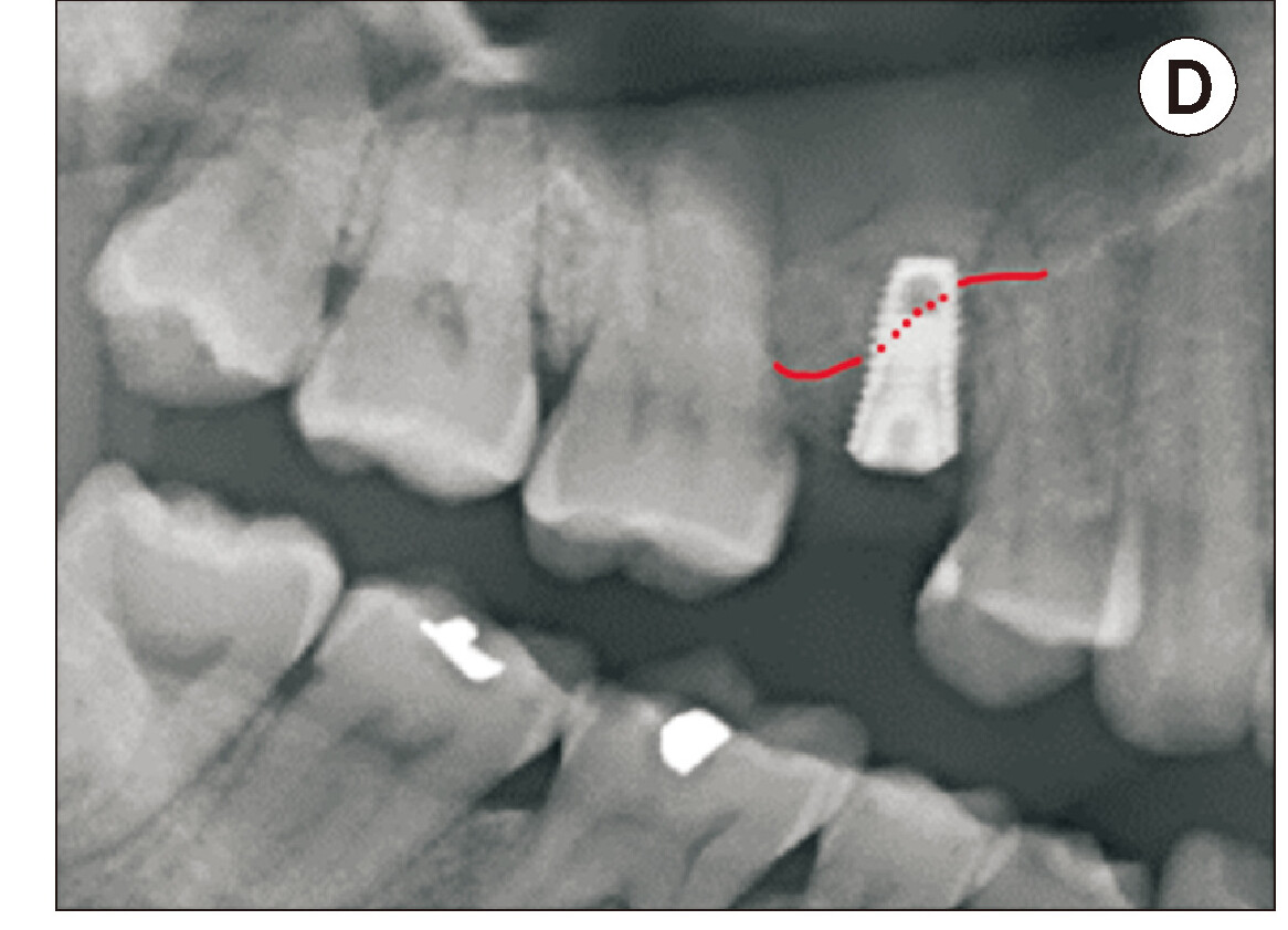
A. Osteotomy prepared to a depth of 3.5 mm. Radiograph with final drill in osteotomy to check for appropriate drilling depth. B. Diagrammatic description of transcrestal sinus floor elevation (tSFE) technique with bone graft placed in osteotomy. C. Use of the ridge spreader at 20 rpm to perform the sinus floor elevation and pack graft material into the sinus. D. Immediate post-operative radiograph showing successful tSFE with dome-shaped radio-opaque bone graft.
Figure 3
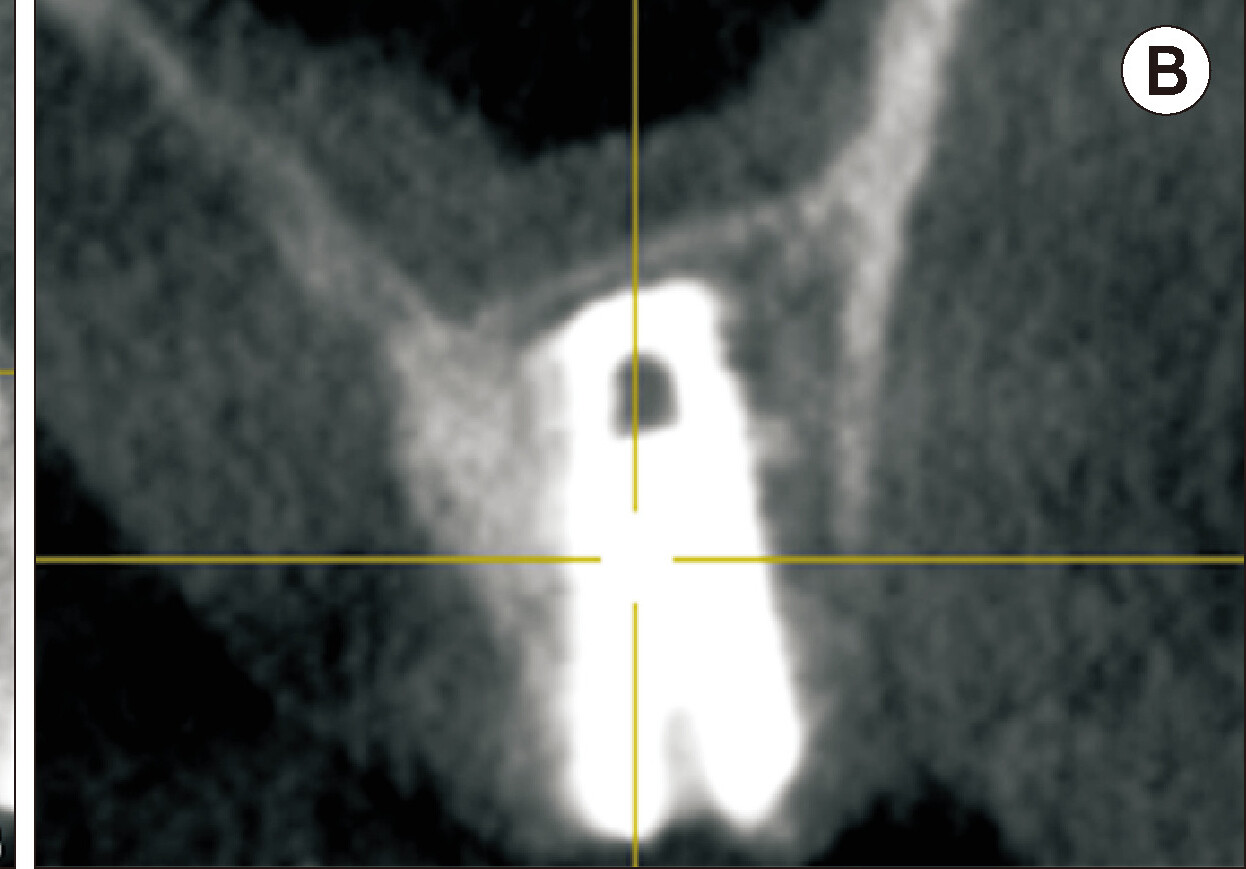
A, B. Six months post-operative cone-beam computed tomography showing successful vertical bone augmentation with the transcrestal sinus floor elevation technique. The apex of the implant is completely surrounded by bone with a layer of cortical bone seen at the sinus floor.
Figure 4
Eight months after the transcrestal sinus floor elevation and simultaneous implant placement, the implant is restored with a screw-retained crown.